Fig. 4-S1
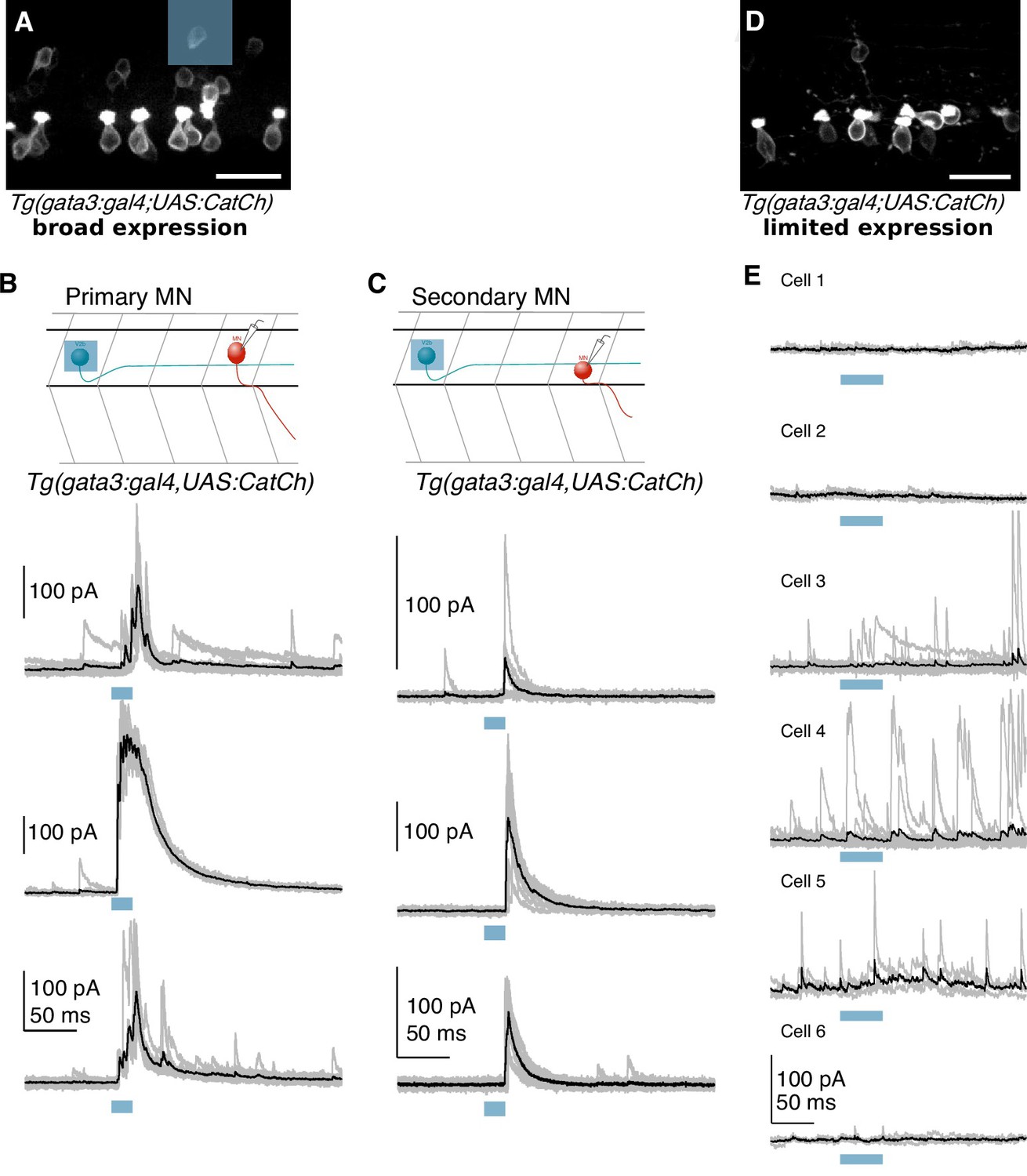
Optogenetically evoked IPSCs originate from V2b neurons, not CSF-cNs.
( A) Confocal projection of a hemisegment of spinal cord in a Tg(gata3:Gal4; UAS:CatCh) animal with broad CatCh expression. A blue square panel shows an example 20 μm x 20 μm DMD stimulation pattern in which 1–2 V2b cells are targeted. Scale bar = 20 μm. ( B) Schematic and whole cell recordings from fast motor neurons in animals with broad CatCh expression, such as in ( A). Shorter stimulation times (20 ms) and targeted illumination, as schematized in ( A), reliably elicit IPSCs in fast motor neurons. ( C) Schematic and recordings from secondary (slow) motor neurons similar to ( D). Light stimulation evoked IPSCs in slow motor neurons in 12/18 neurons. ( D) Confocal image showing limited CatCh expression in a hemisegment of spinal cord in Tg(gata3:Gal4; UAS:CatCh) animals. CatCh is widely expressed in CSF-cN in both animals ( A and D), but from animal to animal, there were variations in the intensity of CatCh expression in V2b neurons. Scale bar = 20 μm. ( E) Whole cell recordings of fast motor neurons in animals with low V2b CatCh expression. Individual traces are shown in gray and averages in black. Blue bar represents the optical stimulation timing. Longer stimulation times (50 ms) and full field illumination were used to maximize IPSC responses in the recorded cell. Motor neurons receive few light-triggered currents in animals with low CatCh expression in V2b neurons. Together this demonstrates that synaptic currents in Figures 5 and 6 originate from V2b and not CSF-cN neurons.

