Image
Figure Caption
Fig. S7
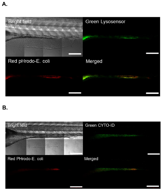
Confocal tiled image with the full zebrafish larvae intestine. (a) The images of bright-field, lysosensor (green), pHrodo-E.coli (red) and merged images of red and green. pHrodo-labelled E. coli were ingested in LRE-enriched region of the intestine. Bar, 200 μm. (b) The images of bright-field, Cyto-ID (green), pHrodo-E.coli (red) and merged images of red and green. Cyto-ID staining is present and not limited to the region with ingested E. coli. Bar, 200 μm.
Acknowledgments
This image is the copyrighted work of the attributed author or publisher, and
ZFIN has permission only to display this image to its users.
Additional permissions should be obtained from the applicable author or publisher of the image.
Full text @ Dis. Model. Mech.

