Image
Figure Caption
Fig. 3
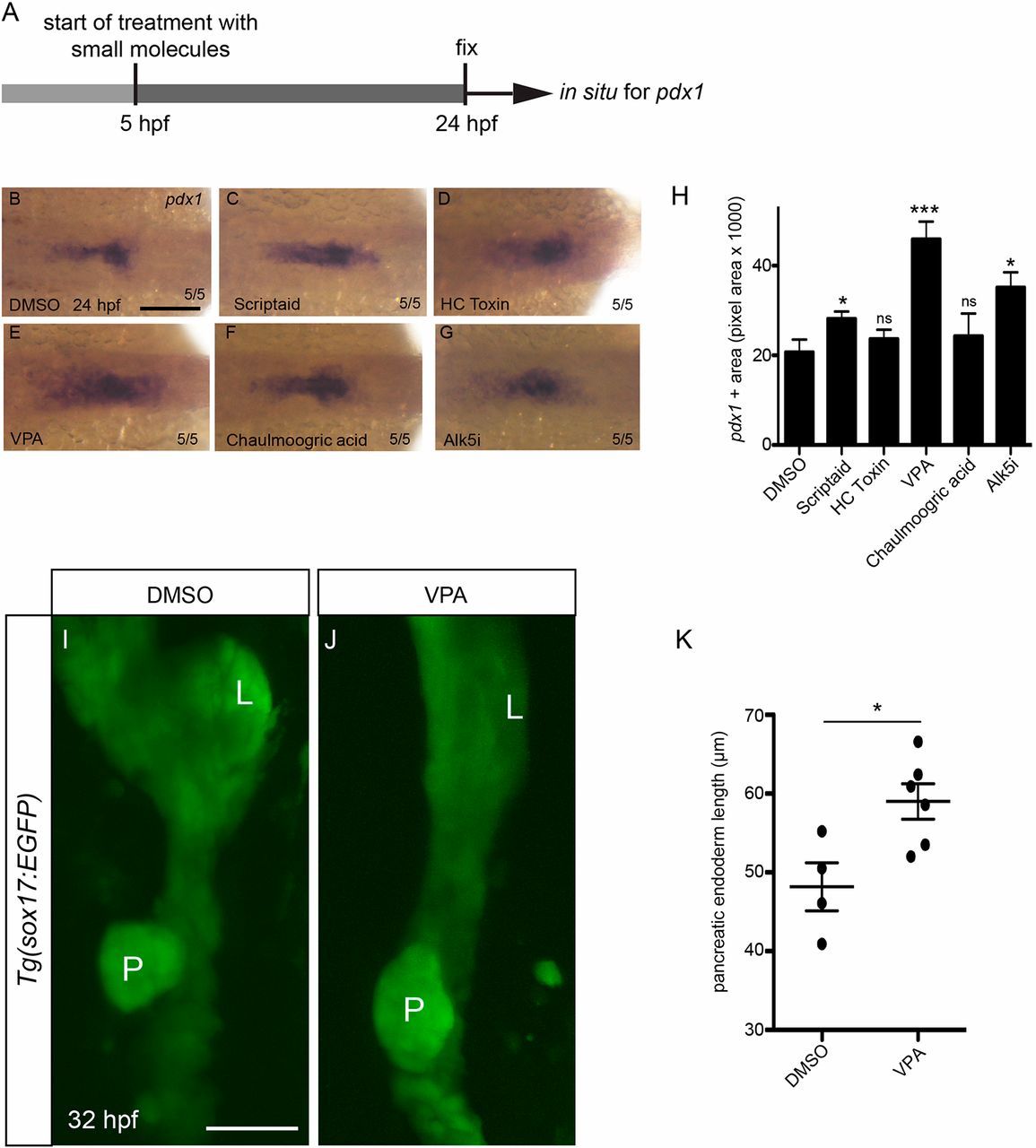
The HDAC inhibitor VPA increases pancreatic endoderm at the expense of hepatic endoderm. (A) Schematic of the experimental set up. (B-G) In situ hybridization on 24 hpf embryos showing pdx1 expression. (H) Scriptaid, VPA and Alk5i treatment lead to an increase in the pdx1-positive area. (I,J) Confocal images of 32 hpf Tg(sox17:EGFP) embryos treated with DMSO (I) as control or VPA (J). L, liver; P, pancreas. (K) VPA treatment leads to an increase in pancreatic endoderm area. ns, not significant. *P≤0.05, ***P≤0.001. Error bars represent s.e.m. Scale bars: 100 µm (B); 20 µm (I).
Acknowledgments
This image is the copyrighted work of the attributed author or publisher, and
ZFIN has permission only to display this image to its users.
Additional permissions should be obtained from the applicable author or publisher of the image.
Full text @ Development

