Fig. 3
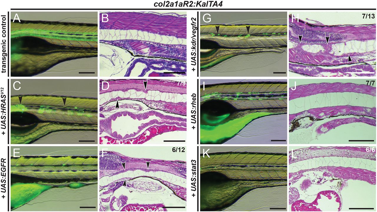
Transient overexpression of RTK genes drives notochord hyperplasia. (A-L) Close-up lateral views of embryo notochords at 5 dpf, with brightfield/fluorescence (left panels) and H&E histology (right panels; different embryos per condition); numbers in D,F,H and J indicate embryos with observed lesions versus phenotypically normal embryos observed in an individual representative experiment, and numbers in L indicate wild-type-looking embryos (n=3 experiments). (A,B) The col2a1aR2:KalTA4 control reference at 5 dpf, expressing UAS:Kaede to fluorescently label the notochord. (C,D) Transient injection of UAS:EGFP-HRASV12 causes localized hyperplasia (arrowheads, C,D) in the notochord. (E,F) Overexpression of human EGFRconsistently causes local hyperplasia in the developing notochord (arrowheads, F). (G,H) Overexpression of zebrafish kdr (encoding the VEGFR2 ortholog) causes strong hyperplasia (arrowheads in G,H, compare with D,F). (I,J) Zebrafish rheb overexpression leads to enlarged vacuoles and no detectable hyperplasia. (K,L) Zebrafish stat3 overexpression leads to no detectable hyperplasia and allows for normal notochord development within 5 dpf. Scale bars: 200 μm.

