
|
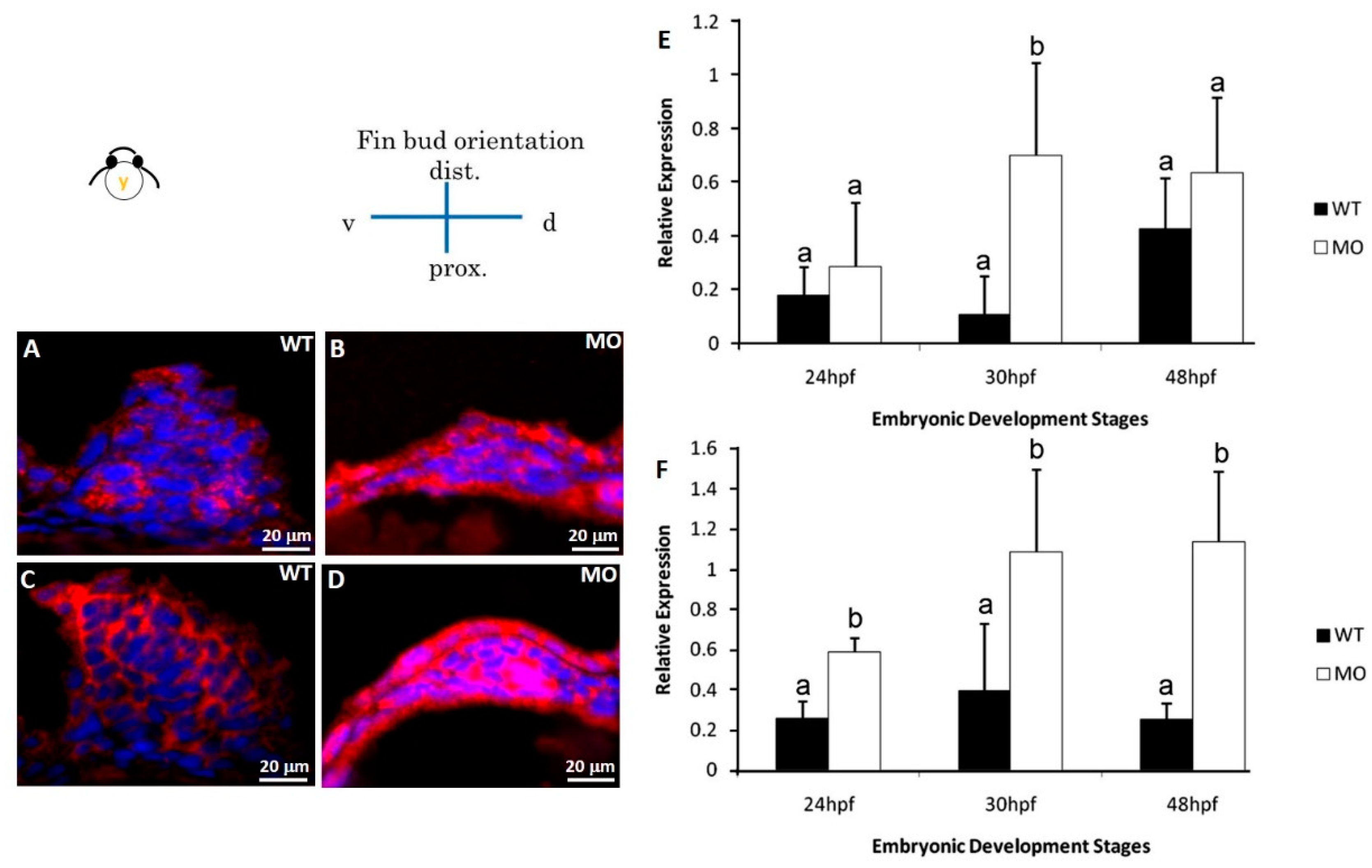
Fig. 7
Immunohistochemical staining of apoptosis-related proteins. Pectoral fins were stained with apoptosis-related (BAD and BCL2) antibodies (red) and counterstained with DAPI (blue) to observe the nucleus. BAD (B) and BCL2 (D) proteins were significantly uninhibited in the tbx5a morphant group compared with those in the Uninjected group (A,C). The embryo anterior is shown on the left. The Bad (E) and Bcl2 (F) expression in the tbx5a morphant group was significantly uninhibited at 24, 30, 36, and 48 hpf (n = 50, triplet) (E). hpf, hours postfertilization; WT, Uninjected group; MO, tbx5a morphant group. a, b: A significant difference was detected by a, b: A significant difference was detected by One-way ANOVA with Duncan’s multiple range test.