
|
Figure 6
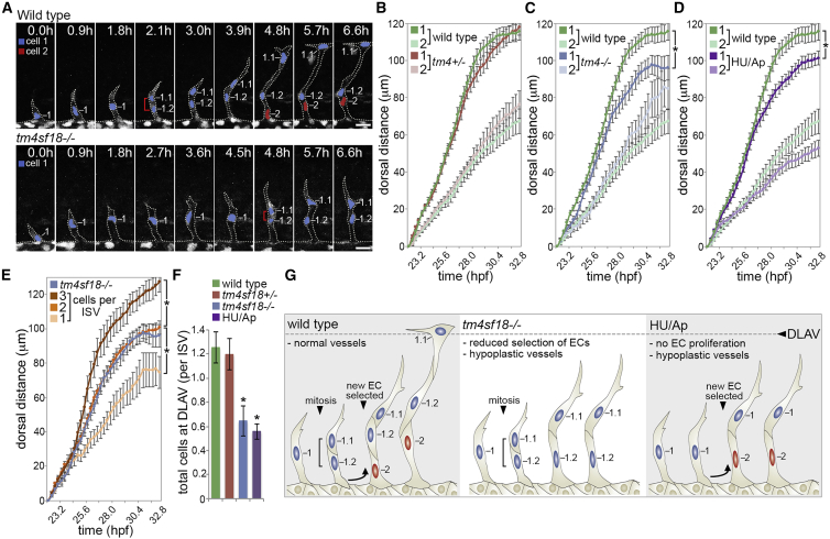
Hypocellular Vessels in
(A) Time-lapse images of sprouting ISVs in WT and
(B–D) Quantification of the dorsal movement of tip (cell 1) or stalk (cell 2) ECs in WT and
(E) Quantification of the dorsal movement of tip ECs in non-proliferating ISVs consisting of 1, 2, and 3 or more ECs and comparison with the motility of tip ECs in
(F) Quantification of the number of ECs that reach the DLAV position in WT,
(G) Illustration of the causes of vessel hypoplasia and phenotypic effect on vessel extension.
Data are means ± SEM. ∗p < 0.05, two-way ANOVA or two-tailed t test. Scale bars, 25 μm.
See also