IMAGE

Fig 5

ID
ZDB-IMAGE-190723-1607
Source
Figures for Oppel et al., 2019
Image
Figure Caption

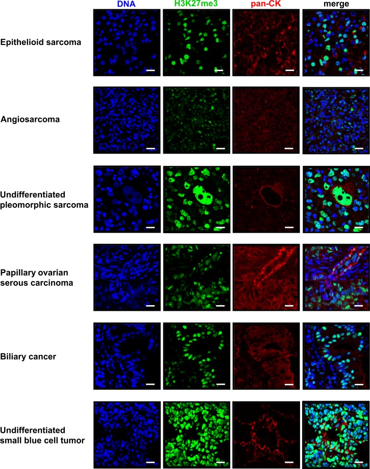
Fig 5 Immunofluorescence staining of tumor types detected in <italic>atrx</italic>+/- zebrafish with <italic>p53/nf1</italic>-deficient background.

This figure shows all tumor types detected in p53-/-, nf1b-/-, nf1a+/-, atrx+/- or p53-/-, nf1b-/-, nf1a+/+, atrx+/- fish that were not observed in atrx-wildtype siblings. For each tumor a section stained by indirect immunofluorescence for H3K27me3 (green) and pan-cytokeratin (red) is shown. The DNA is visualized with Hoechst (blue); scale bars: 10μm.

Acknowledgments
This image is the copyrighted work of the attributed author or publisher, and ZFIN has permission only to display this image to its users. Additional permissions should be obtained from the applicable author or publisher of the image. Full text @ PLoS Genet.