Fig. S3
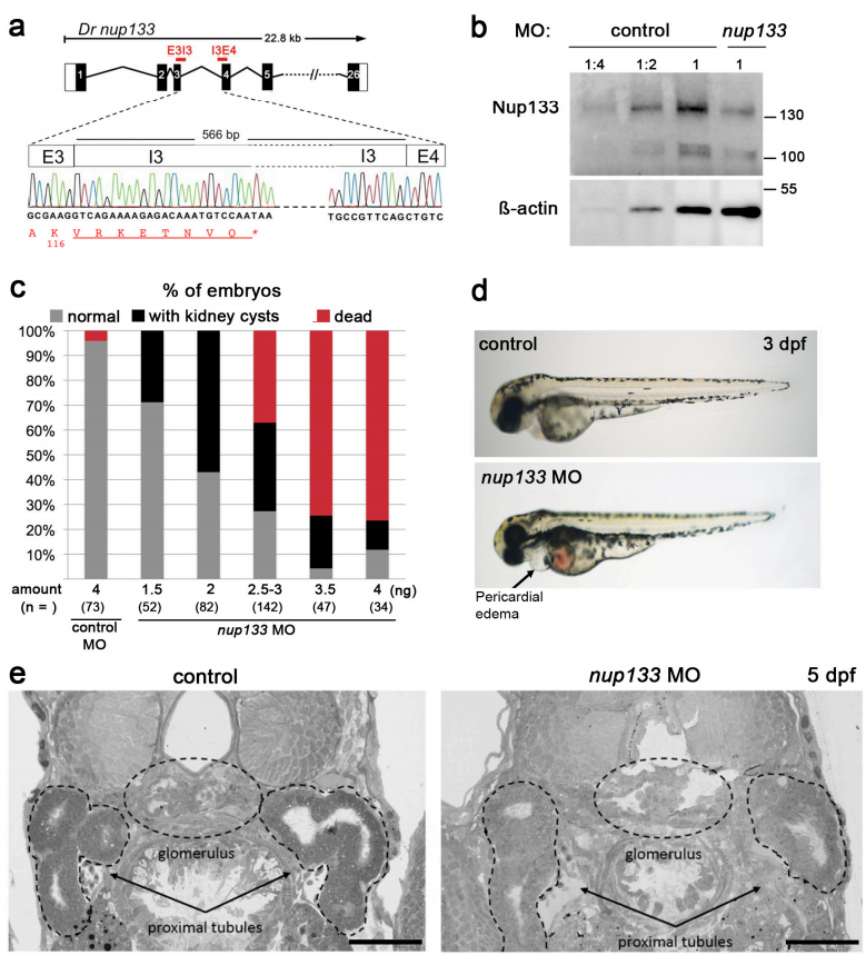
Retention of intron 3 in nup133 MO injected embryos and morphology of 3 and 5 dpf control and nup133 MO larvae. (a) Exon structure of Danio rerio (Dr) nup133 around the binding sites of the E3I3 and I3E4 splice morpholinos. The size of intron 3 is indicated. Sequencing of the additional RT-PCR product in the nup133MO embryos shows retention of intron 3. The end of the predicted aa sequence of the corresponding truncated protein is indicated in red, under the DNA sequence. Residues encoded by the intron are underlined. (b) Extracts from 24 hpf control and nup133 MO embryos were analyzed by western blot using anti-Nup133 and anti ß-actin antibodies (used as loading control). Dilutions of the control samples (1:2 and 1:4) were also loaded to better appreciate the decrease of Nup133 protein level. (c) Relative proportions of 3dpf embryos with kidney cysts or dead upon injection of the indicated amounts of control or of each nup133 MO. For each condition, the total number of embryos analyzed is indicated (n=). (d) Gross morphology of 3 dpf control and nup133 MO larvae. Note the pericardial edema in nup133 MO larvae (arrow). (e) Toluidine blue stained sections of 5 dpf uninjected control (left panel) and nup133 MO injected larvae (right panel) showing the glomerulus and the proximal tubules (indicated with dotted lines). Note the cystic dilation of the glomerulus in the nup133 MO larvae that is not associated with a major dilatation of the proximal tubules. Scale bars 50 µm.

