Fig. 6
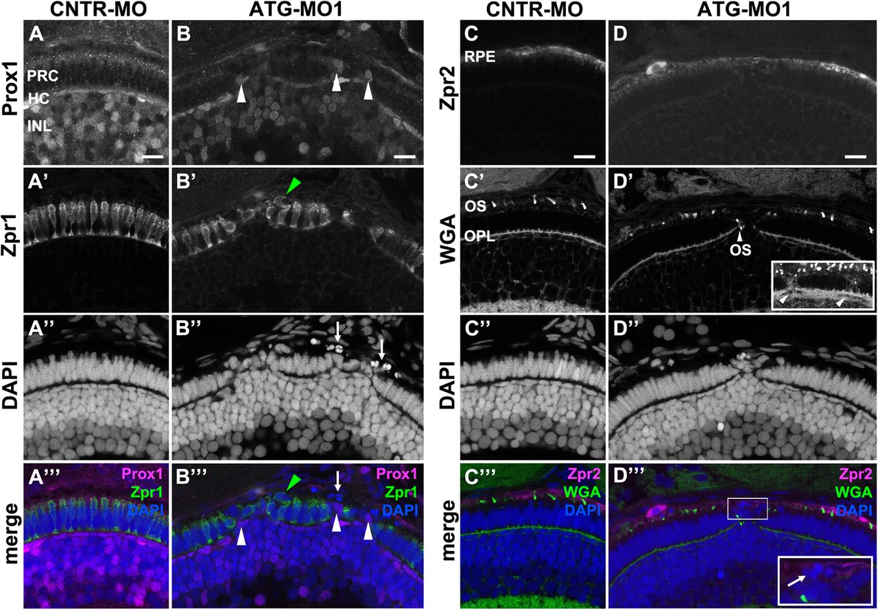
Knockdown of Lgl2 in pen/lgl2 clutches affects the organization of the outer and inner nuclear layers.Immunostaining of transverse retinal sections at 3 dpf of MO-injected pen/lgl2 clutches. (A–A′′′,C–C′′′) pen/lgl2 clutch injected with CNTR-MO and (B–B′′′,D–D′′′) pen/lgl2 clutch injected with ATG-MO1. (A–B′′′) Prox1- (A,B) and Zpr1-(A′,B′) positive cells are well separated from each other in control retinas (A–A′′′) but mix in Lgl2 morphants (B–B′′′). White arrowheads in B and B′′′ denote Prox1-positive cells that have been apically displaced. Green arrowheads in B′ and B′′′ mark an apically displaced PRC. Arrows in B″ and B′′′ mark pyknotic nuclei apical to the photoreceptor layer. (C–D′′′) Zpr2-positive RPE cells (C,D) in MO-injected pen/lgl2 clutches (D–D′′′) surround nuclei apical to the PRC layer. WGA staining (C′,D′) shows that in Lgl2 morphants, disorganized PRCs have outer segments (OS), but these can be found next to the OPL (arrowhead in D′). Projection of multiple optical planes shows breakages in the plane of OPL (inset in D′, arrowheads). Inset in D′′′ shows pyknotic nuclei (arrow) that appear to be surrounded by the Zpr2 signal. PRC, photoreceptor cells; HC, horizontal cells; INL, inner nuclear layer; OPL, outer plexiform layer. Scale bars: 10 µm.

