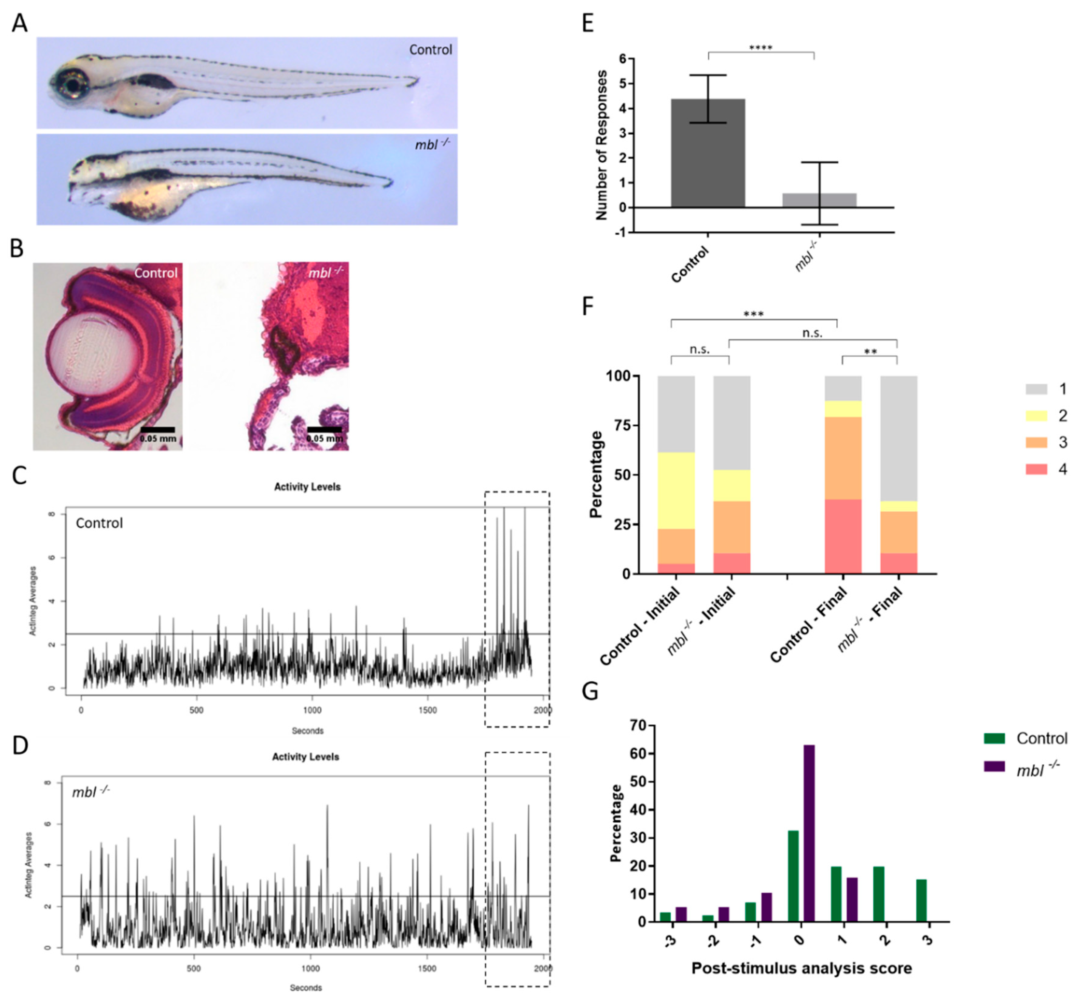
Fig. 3
Utilizing the eyeless masterblind (mbl) mutant as a negative control for VIZN and OMR. (A) 6 days post-fertilization control (top) and mbl−/−mutants (bottom); (B) Hematoxylin and eosin staining larvae in (A). Control larvae display normal optic structures and retinal lamination while homozygous mbl−/− mutants lack eyes; (C) Activity profile of control and, (D) mbl−/−; (E) VIZN analysis between control (n = 39) and mbl−/− (n = 28) (Mann–Whitney, p-value **** < 0.0001); (F) OMR analysis of larvae plotted as a bar graph which shows the shifts in the population between the initial and final positions (Bowker’s test of symmetry, p-value *** = 0.0004; Wilcoxon–Mann–Whitney, p-value ** = 0.0019); (G) the post-stimulus analysis which takes the difference between the final and initial position to show positional changes in individual fish. The same larvae were used for all assays. Scale bars in (B): 0.05 mm. n.s., not significant.

