
|
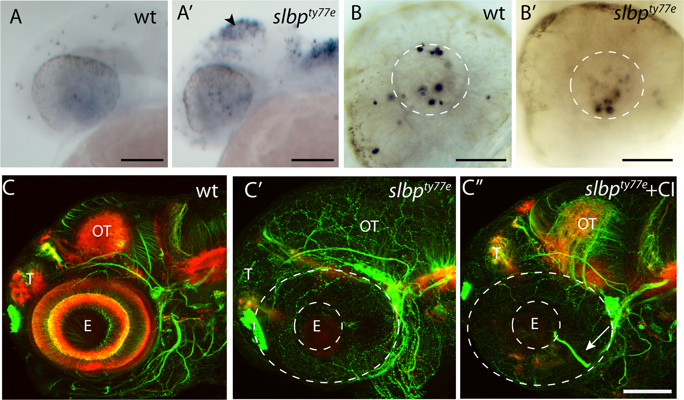
Fig. 4
(A-B’) Lateral view of 26hpf wild type (A,B) and slbpty77e (A’,B’) heads and eyes stained with TUNEL. Apoptosis is prominent in the tectum (arrowhead) and dorsal hindbrain of the mutant (A’) but not within the eye (B’). The white lines outline the lens within which there is apoptosis in both the wildtype and mutant eyes. (C-C”) Lateral view of 72hpf untreated wildtpe (C), untreated slbpty77e (C’) and (C”) caspase inhibitor treated slbpty77e heads labelled with anti-acetylated tubulin (axons, green) and anti-SV2 (neuropil, red). Arrow shows aberrant retinal ganglion axons in the mutant eye. Abbreviations: AC, anterior commisure; E, eye; OT, optic tectum; T, telencephalon. Scale bar: (A-A’,C-C”) 100μm; (B-B’) 50μm.