Image
Figure Caption
Fig. 2
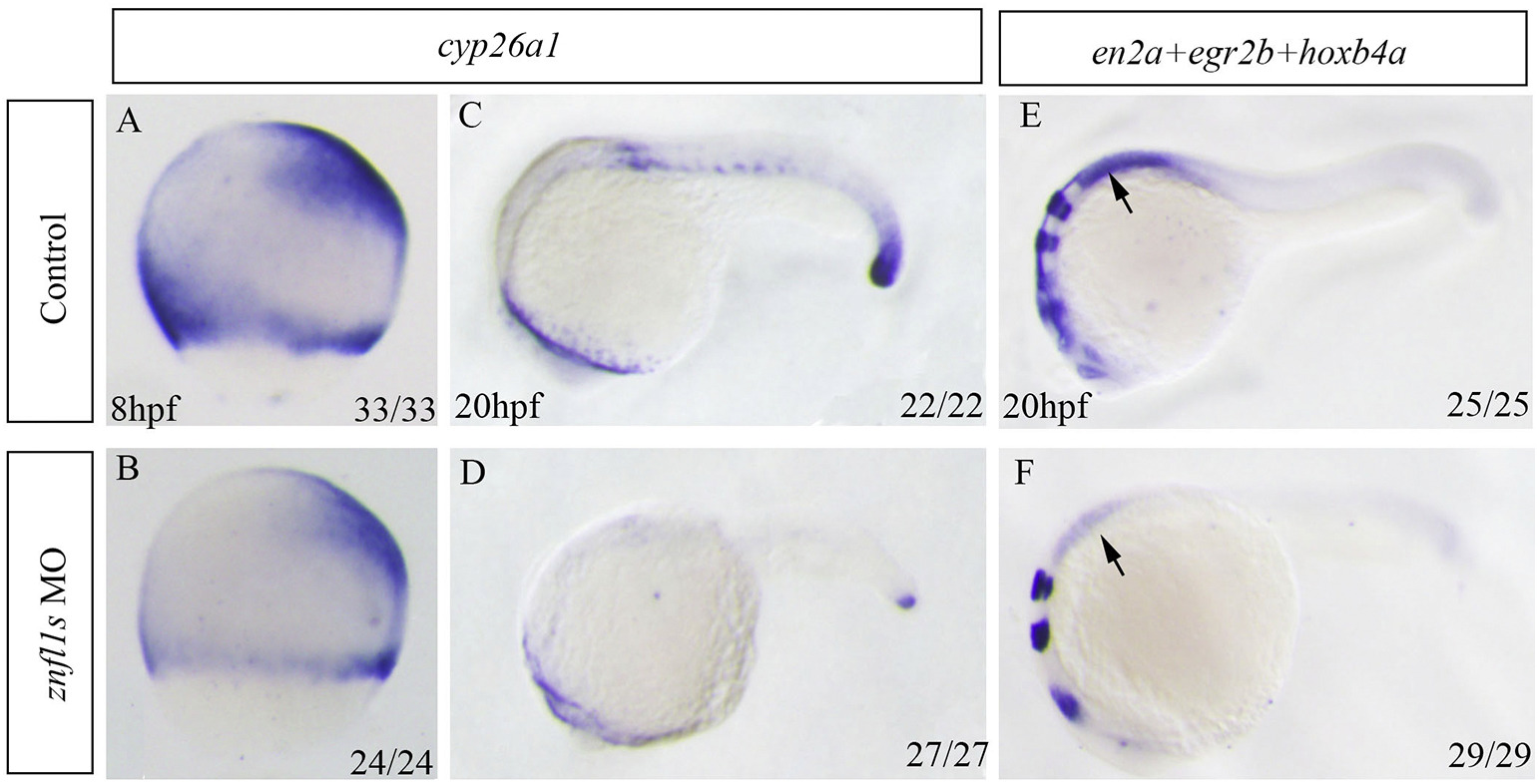
Knocking down znfl1s reduces RA signaling in zebrafishembryos. Whole mount in situ hybridized embryos are positioned animal pole top (8 hpf, A–B) or anterior left (20 hpf, C–F). The expression of cyp26a1 was examined in controls (A, C) and znfl1s morphants (B) at 8 hpf or 20 hpf (D). The expressions of en2a, egr2b and hoxb4a in control embryos (E) and znfl1smorphants (F). All embryos were positioned in lateral view. The arrow points to the expression of hoxb4a in E–F.
Figure Data
Acknowledgments
This image is the copyrighted work of the attributed author or publisher, and
ZFIN has permission only to display this image to its users.
Additional permissions should be obtained from the applicable author or publisher of the image.
Reprinted from Mechanisms of Development, 155, Li, J., Zhao, Y., He, L., Huang, Y., Yang, X., Yu, L., Zhao, Q., Dong, X., Znfl1s are essential for patterning the anterior-posterior axis of zebrafish posterior hindbrain by acting as direct target genes of retinoic acid, 27-33, Copyright (2018) with permission from Elsevier. Full text @ Mech. Dev.

