
Image
|
Figure Caption
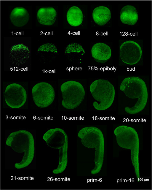
Fig. 1 Distribution of the CTCF protein through development. Whole-mount immunoflourescence staining of zebrafish embryos, from the 1-cell to the prim-16 stages (staging according to Kimmel et al., 1995), encompassing the first 30 h of development.
Figure Data
Acknowledgments
This image is the copyrighted work of the attributed author or publisher, and
ZFIN has permission only to display this image to its users.
Additional permissions should be obtained from the applicable author or publisher of the image.
Reprinted from Mechanisms of Development, 154, Carmona-Aldana, F., Zampedri, C., Suaste-Olmos, F., Murillo-de-Ozores, A., Guerrero, G., Arzate-Mejía, R., Maldonado, E., Navarro, R., Chimal-Monroy, J., Recillas-Targa, F., CTCF knockout reveals an essential role for this protein during the zebrafish development, 51-59, Copyright (2018) with permission from Elsevier. Full text @ Mech. Dev.