
|
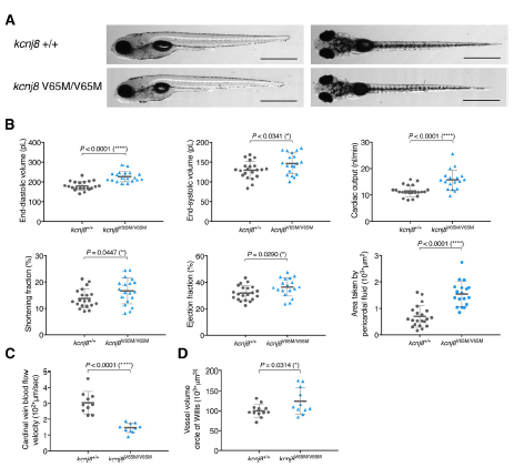
Fig. S7
Homozygous kcnj8V65M/V65M mutation induces CS-related cardiac anomalies and cerebral vasodilation in zebrafish embryos. (A) Representative images illustrating the morphology of 5 dpf wild-type and kcnj8V65M/V65M mutants as seen from a left lateral (top) and dorsal view (bottom). (B) Quantification of cardiac function using individual characteristic confocal sections from a time series of the embryonic cardiac cycle at 5 dpf. Pericardial edema was quantified by measuring pericardial area using striking morphological landmarks indicated by white boxes. (C) Tracking of individual red blood cells (RBCs) measuring blood flow velocity in the cardinal vein. RBCs were tracked for 10 frames using ImageJ (NIH) and the plugin MTrackJ(35). (D) Quantification of vascular dilations in a Tg(kdrl:GFP) background. 3D reconstruction of vascular structure in Imaris was used to calculate vessel volume. kcnj8+/+ controls are the same as in Fig.2B-D. For all graphs, significance was determined by two-tailed unpaired Student's t test or Mann–Whitney two-tailed U test: * p≤0.05; ** p≤0.01; *** p≤0.001; **** p≤0.0001. The black horizontal bar indicates the mean value for each condition. Sample size, kcnj8+/+, n=21; kcnj8V65M/V65M, n=19 in B, kcnj8+/+, n=10; kcnj8V65M/V65M, n=10 in C, kcnj8+/+, n=20; kcnj8V65M/V65M, n=11 in D. Scale bars, 1 mm A. All embryos analyzed originated from group matings of adult zebrafish.