Fig. 7
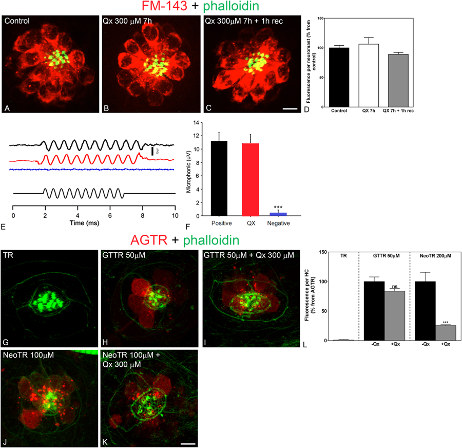
Treatment with Qx does not abolish mechanotransduction channel activity. 5dpf wild type larvae were incubated in E3 media (A), Qx 300 μM for 7 hours and immediately assayed for FM1-43 uptake (B) or Qx, 300 µM, recovered for 1 hour and then treated with the dye (C). Quantification of the fluorescent intensity per neuromast (D) was expressed as percentage from controls. No significant differences were observed between control and treated animals (unpaired Student’s t-test). (E,F) Microphonic potentials from animals treated with vehicle (black) or 300 μM of Qx (red). Pcdh15a mutants were used as a negative control (blue). Microphonic responses are represented as mean +/− SD. Only the Pcdh15a mutants showed a significant decrease in microphonic potentials compared to controls (unpaired Student’s t-test). (G–L) 5dpf zebrafish were incubated with hydrolyzed Texas Red (TR, G) for 1 hour, gentamicin-conjugated Texas Red (GTTR, H) 50 µM for 1 hour, Qx 300 µM for 8 hours +GTTR 50 µM for 1 hour before the end of the experiment (I), neomycin-conjugated Texas Red (NeoTR, J) 100 µM for 30 min or with Qx 300 µM for 8 hours +NeoTR 100 µM for 30 min before the end of the experiment (K). (L) The fluorescence intensity incorporated per HC was expressed as a percentage from the corresponding aminoglycoside-Texas Red (AGTR) incubation treatment and represented as mean +/− SEM. Unpaired Student’s t-test. ***p < 0.001. Student’s t-test versus corresponding aminoglycoside-only treatment. Scale bar: (A–C) 6 μm, (G–K) 5 µm. Data were taken from at least 10 animals and 3 experiments runs. For microphonic potentials 8–9 animals were analyzed per treatment.

