
|
Fig. 3
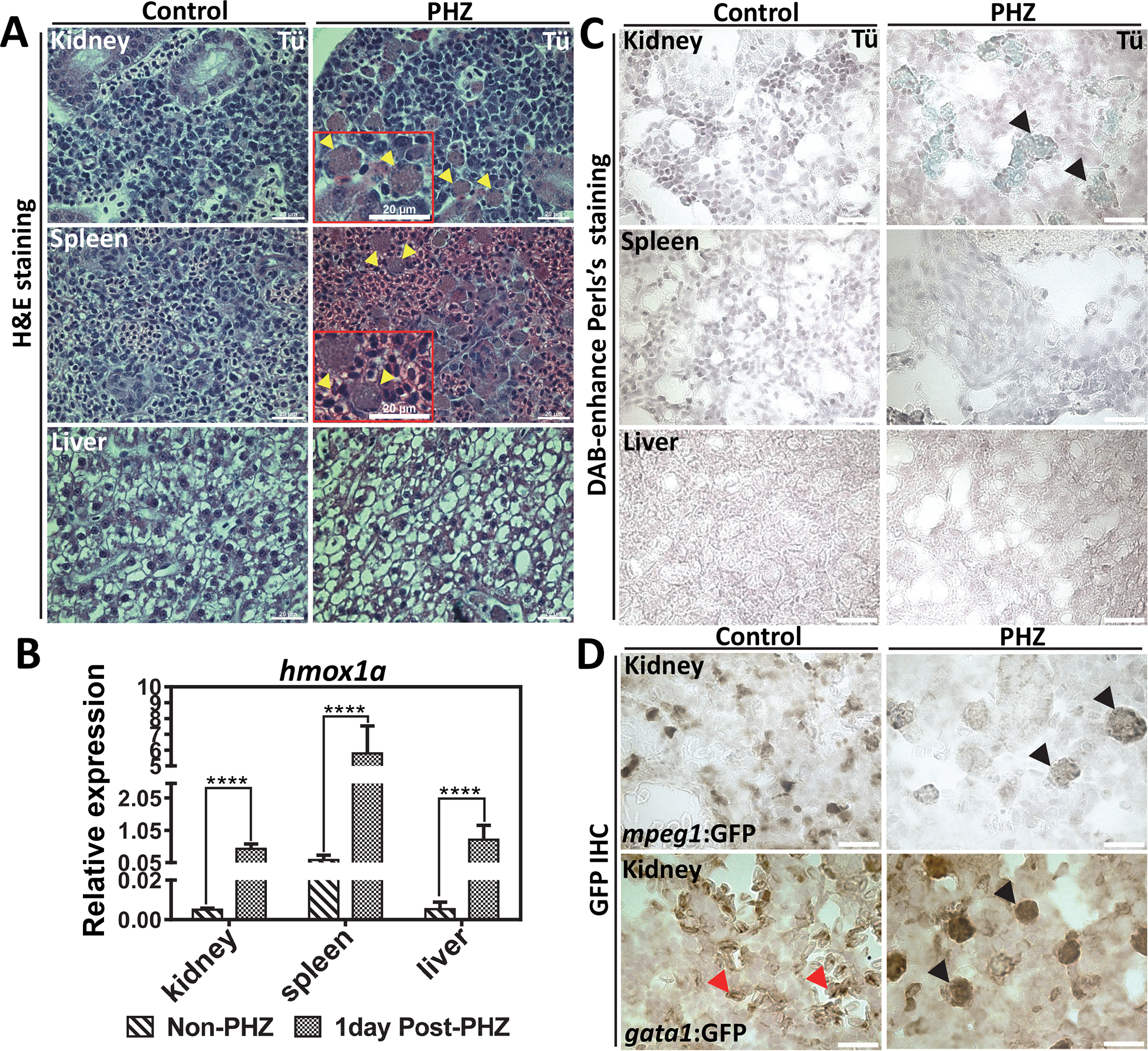
The zebrafish kidney is responsible for heme-iron recycling during EP.
(A) H&E staining of kidney, spleen and liver sections from adult WT zebrafish with control (non-PHZ treated) and 1 day post PHZ-treatment samples. Cells undergoing erythrophagocytosis are indicated as yellow arrows. (B) qRT-PCR of hmox1a mRNA expression in the kidney, spleen and liver from control (non-PHZ treated) and PHZ treated adult zebrafish at 1day after treatment. Three adult zebrafish were pooled as one cohort, and three cohorts were repeated as biological triplicates. * p<0.05. (C) Perl’s Prussian blue iron staining on sections of kidney, spleen and liver from adult Tü WT zebrafish with control (non-PHZ treated) and 1 day post PHZ-treatment samples. Positive iron staining is indicated in kidney macrophages as showed by black arrows. (D) GFP IHC of kidney sections from adult transgenic zebrafish gata1:gfp and mpeg1:gfp with control (non-PHZ) and 1 day post PHZ-treatment. RBCs are indicated with red arrows and macrophages with black arrows.