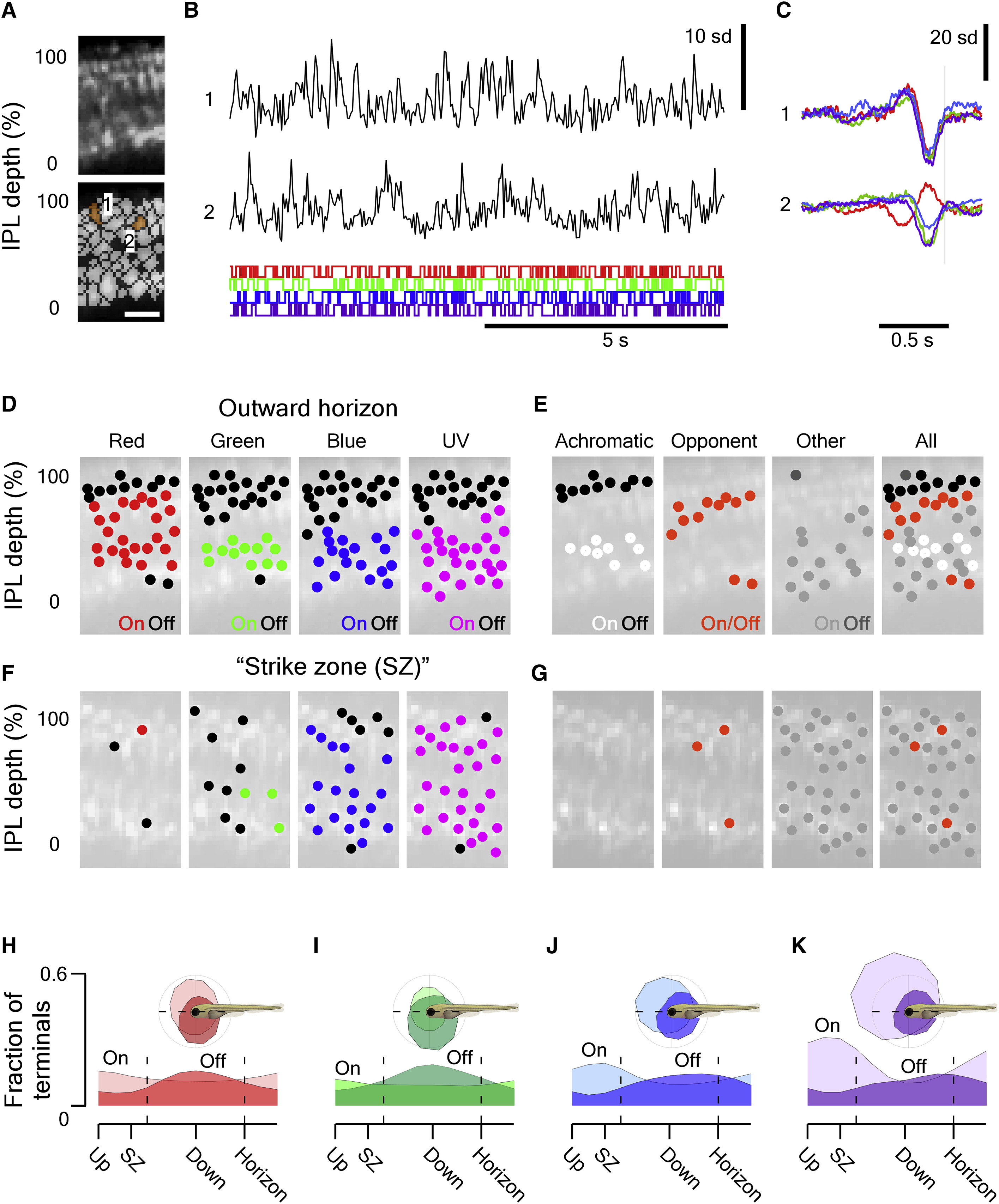
Fig. 3
Surveying Inner Retinal Chromatic Responses In Vivo
(A) 2-photon scan field (32 × 64 pixels; 15.625 Hz) of a nasal IPL section (outward horizon) in a Tg(−1.8ctbp2:SyGCaMP6) larvae for simultaneous recording of light-driven calcium responses across the entire IPL depth at single-terminal resolution (top) and regions of interest (ROIs) (bottom). The scale bar represents 5 μm.
(B) Example of calcium responses to tetrachromatic binary white noise stimulation (12.8 Hz; STAR Methods) of two ROIs highlighted in (A).
(C) Tetrachromatic linear filters (“kernels”) recovered by reverse correlation of each ROI’s response with the noise stimulus (B). The color code indicates the stimulus channel (R, G, B, U; cf. Figure S3A).
(D) For each stimulus channel, we classified each ROI’s kernel as either “on” (in red, green, blue, or purple), “off” (black), or non-responding (no marker) and plotted each response over the anatomical scan image (STAR Methods).
(E) By comparison across the four stimulus channels, we then classified each ROI as either achromatic off (R+G+B+U off, black) or on (R+G+B+U on, white), Color opponent (any opposite polarity responses in a single ROI, orange) or “other” (gray) and again plotted each ROI across the IPL to reveal clear chromatic and achromatic layering in this scan is shown.
(F and G) As (D) and (E), respectively, but for a scan taken in the temporo-ventral retina, which surveys the world in front of the animal just above the visual horizon. This zone is critical for prey capture and was thus dubbed “strike zone”.
(H–K) Distribution of all on and off responses per stimulus channel (H, red; I, green; J, blue; and K, UV) based on n = 4,099/6,565 ROIs that passed a minimum quality criterion (STAR Methods) sampled from across the entire sagittal plane (115 scans, 12 fish).
See also Figure S3.

