
|
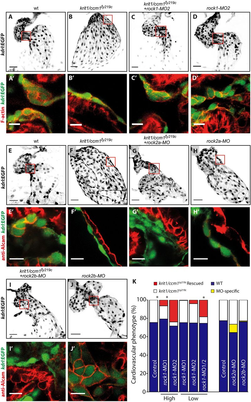
Fig. 5
ROCK1 silencing in ccm1 zebrafish mutant embryos rescues cardiac morphogenesis. (A–J) Endocardial cells, marked by Tg(kdrl:EGFP) (inverted images) at 48 hpf. Details with single confocal plane section of the atrioventricular canal (AVC) are shown in A′–J′. Scale bars: 30 µm (main images); 10 µm (detailed images). (K) Quantification of the number of embryos that showed a phenotypic rescue of the krit1/ccm1ty219c cardiac phenotype at 48 hpf. Embryos from krit1/ccm1ty219c/+ incrosses were 75% phenotypically WT (blue bars), while 25% showed krit1/ccm1ty219c mutant phenotype of cardiac ballooning (white bars). Embryos from krit1/ccm1ty219c/+ incrosses that were injected with high doses of rock1-MO1 and -MO2 showed a rescue of the cardiac ballooning (red), whereas embryos injected with low MO doses failed to show significant numbers with rescue. However, when combined, low doses of rock1-MO1 and -MO2 rescued the cardiac ballooning phenotype in krit1/ccm1ty219c mutant embryos as efficiently as single high dose MOs. Total numbers quantified, from a set of three independent zebrafish cohorts, are n=195 for control embryos; rock1-MO1 injected at high dose, n=232; rock1-MO2 at high dose, n=126; rock1-MO1 at low dose, n=167; rock1-MO2 at low dose, n=148; and combined rock1-MO1 and MO2 at low dose, n=134. *P<0.05 (two-way ANOVA with Dunett's multiple comparisons post-test). Embryos from krit1/ccm1ty219c/+ incrosses that were injected with either rock2a-MO1 or rock2b-MO2 did not rescue the cardiac ballooning phenotype, however, a fraction of the sibling population show MO-specific defects that are similar to the krit1/ccm1ty219c phenotype. Control for rock2a-MO, n=230; rock2a-MO, n=265; control for rock2b-MO, n=526; rock2b-MO, n=317.