Image
Figure Caption
Fig. S4
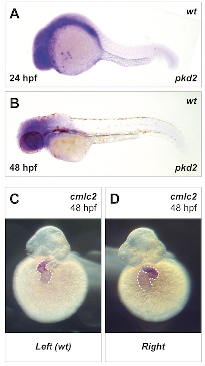
Loss of pkd2 expression in zebrafish caused randomization of left–right patterning.
(A) In situ hybridization of pkd2 mRNA in wild-type zebrafish 24 (B) and 48 hpf. (C,D) Left–right asymmetry was visualized by in situ hybridization for cmlc2 to evaluate heart looping. Knockdown of pkd2 expression caused a randomization of heart looping [25]. cmlc2, cardiac myosin light chain 2; hpf, hours post fertilization.
Acknowledgments
This image is the copyrighted work of the attributed author or publisher, and
ZFIN has permission only to display this image to its users.
Additional permissions should be obtained from the applicable author or publisher of the image.
Full text @ PLoS Biol.

