Fig. S16
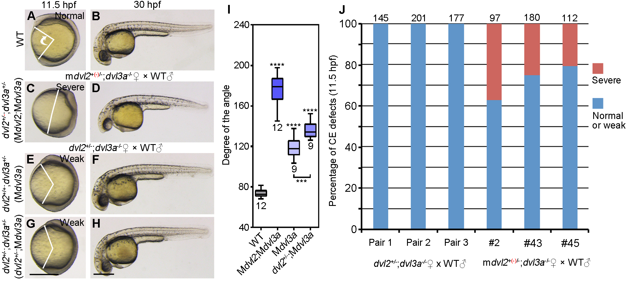
Maternal contribution of Dvl2 and Dvl3a in axis extension.
The embryos were imaged at 11.5 hpf and 30 hpf, followed by genotyping, and the extent of axis extension defect was reflected by the angle between the anterior end and the posterior end at 11.5 hpf. (A, B) WT embryos. (C, D) Mdvl2;Mdvl3a mutants from crosses between female mdvl2+(-)/-;dvl3a-/- fish and male WT fish were genotyped for the presence of a novel indel (red) along with a WT allele in the dvl2 locus. They have dvl2 and dvl3a heterozygous mutations. (E, H) The offspring with the two possible genotypes (dvl2+/+;dvl3a+/- and dvl2+/-;dvl3a+/-; the effects of these mutations are indicated in parenthesis), derived from a cross between female dvl2+/-;dvl3a-/- fish and male WT fish, are maternal mutants for dvl3a, with a reduced dosage of maternal dvl2 in both cases, despite of the genotype. (I) Statistical analysis of the extent of axis extension delay in three types of maternal mutants. Bars represent the mean ± s.d. from indicated numbers of embryos, and asterisks above the bars show significance with respect to WT embryos (***, P<0.001; ****, P<0.0001). (J) Quantitative analysis of defective axis extension at 11.5 hpf. Each type of cross was done using three independent fish pairs, and total numbers of embryos analyzed are indicated on the top of each column. Subjective measures of axis extension defect are shown on the embryos at 11.5 hpf. Scale bar: (A, C, E, G) 400 μm; (B, D, F, H) 400 μm.

