Fig. 7
Tdrd6a Stimulates Buc-eGFP Mobility In Vivo
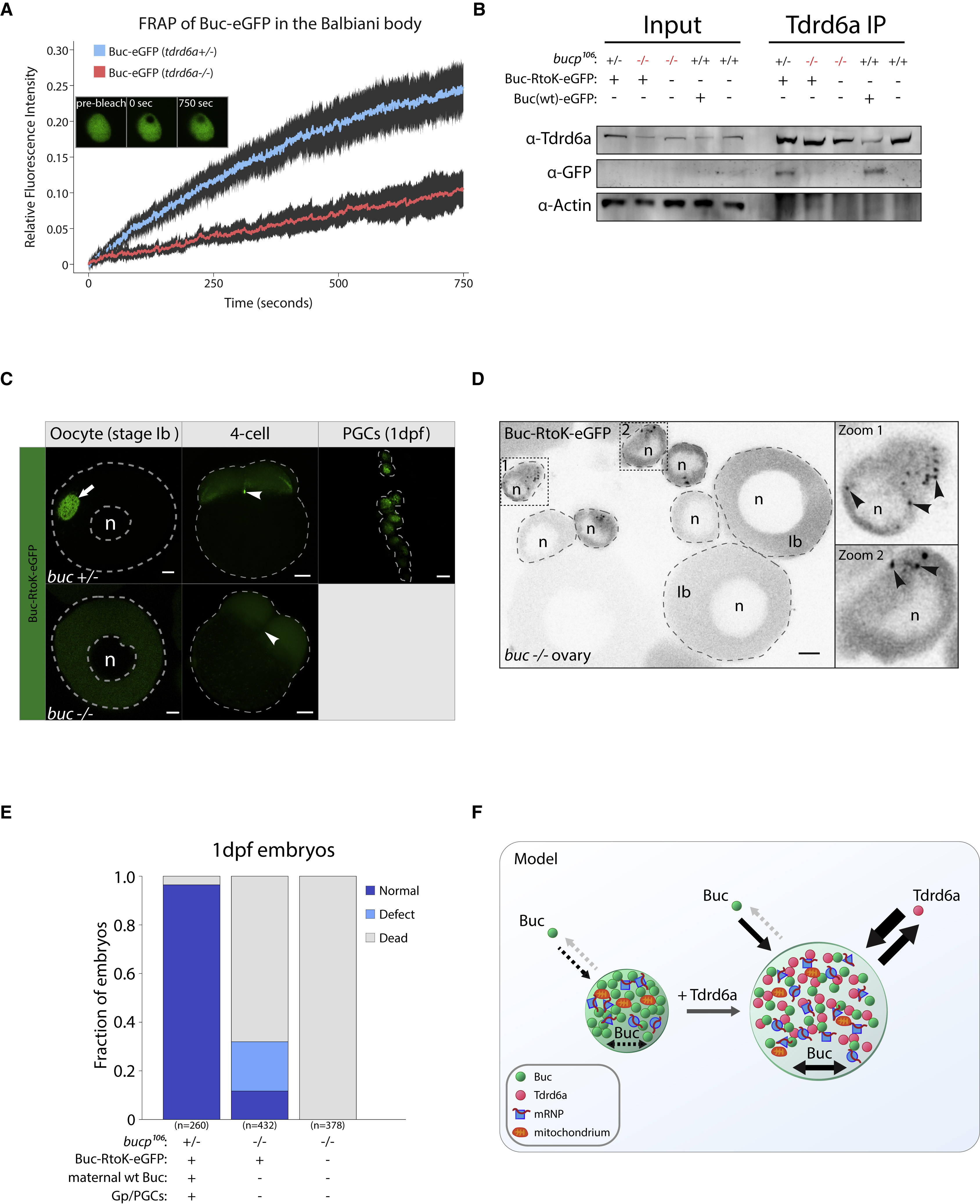
(A) FRAP recovery curves of Buc-eGFP in tdrd6a heterozygous or mutant Balbiani bodies. Fluorescence intensity is the calculated fraction of the pre-bleach intensity and plotted with a 95% confidence interval.
(B) Tdrd6a IPs probed for the indicated proteins by western blot. Bucp106 = buc loss-of-function allele. Note that Buc-eGFP is typically very hard to detect in total lysates.
(C) Localization of Buc-RtoK-eGFP in the buc+/− and buc−/− background. Arrow indicates Bb, arrowheads indicate Gp (buc+/−) or where Gp should be (buc−/−). Scale bars for oocyte and 1dpf, 10 μm. Scale bar for 4-cell, 100 μm. n = nucleus.
(D) Overview of buc−/− ovary (whole mount) positive for Buc-RtoK-eGFP. Zooms 1 and 2 are examples of stage-I oocytes ø < ∼30 μm, containing small Buc-RtoK-positive granules (arrowheads). These granules are never detected in stage-Ib oocytes, where Buc-RtoK is diffusely cytoplasmic. Scale bar, 10 μm, n = nucleus.
(E) Quantification of progeny viability at 1 dpf spawned by mothers with background as indicated, crossed with wt males.
(F) Model of Buc-containing granules, with or without Tdrd6a. Arrows indicate movement in and out of the structure or mobility within the structure itself.
See also Figure S7.
Reprinted from Developmental Cell, 46, Roovers, E.F., Kaaij, L.J.T., Redl, S., Bronkhorst, A.W., Wiebrands, K., de Jesus Domingues, A.M., Huang, H.Y., Han, C.T., Riemer, S., Dosch, R., Salvenmoser, W., Grün, D., Butter, F., van Oudenaarden, A., Ketting, R.F., Tdrd6a Regulates the Aggregation of Buc into Functional Subcellular Compartments that Drive Germ Cell Specification, 285-301.e9, Copyright (2018) with permission from Elsevier. Full text @ Dev. Cell

