
|
Fig. 2
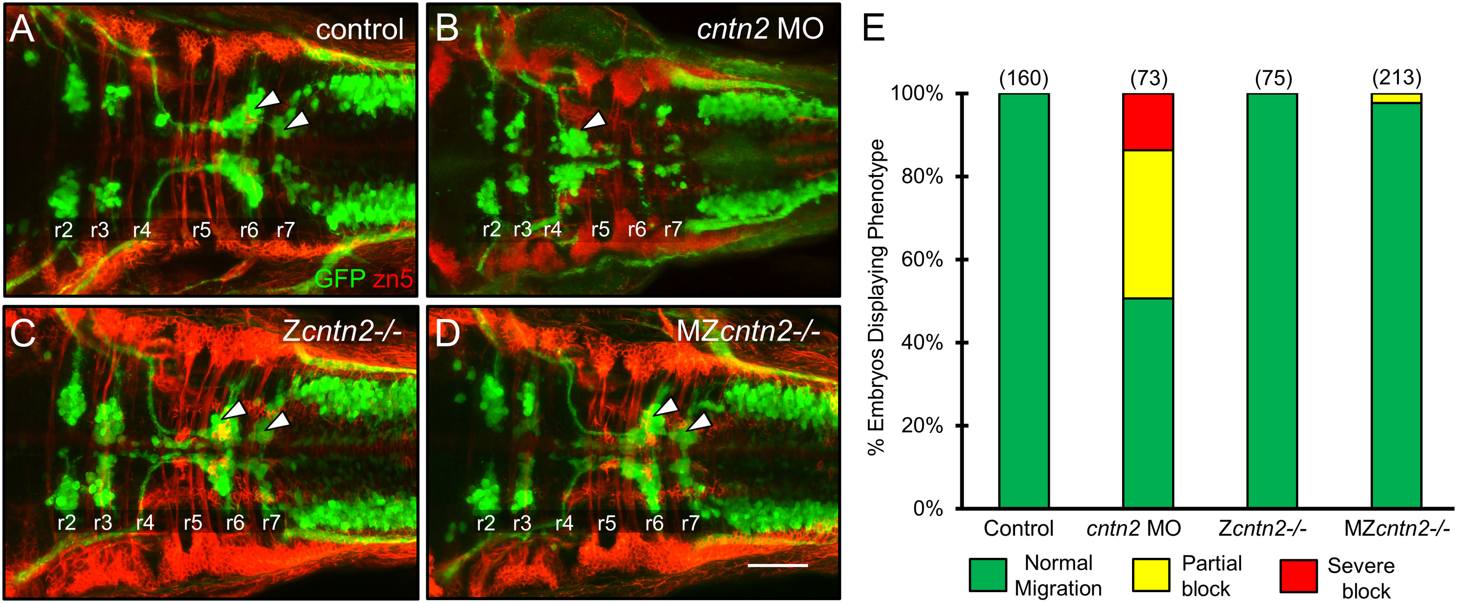
FBM neuron migration is affected in cntn2 morphants but not in cntn2 mutants
Panels A-D show dorsal views of the hindbrain with anterior to the left. Tg(isl1:gfp) embryos were fixed at 48 hpf, and processed for immunohistochemistry with zn5 antibody (red) to label hindbrain commissural neurons and axons at rhombomere boundaries, and anti-GFP antibody (green) to label FBM neurons (arrowheads). (A) FBM neurons (arrowheads) migrate normally into r6 and r7 in an uninjected embryo. (B) FBM neurons largely fail to migrate out of r4 in a cntn2 MO-injected embryo. (C, D) FBM neurons migrate normally in zygotic mutant (Zcntn2−/−) (C), and maternal-zygotic mutant (MZcntn2−/−) (D) embryos. Scale bar in D, 50 μm for A–D. (E) Quantification of FBM neuron migration defects. Number in parenthesis denotes number of embryos. Data are from 3 to 4 experiments. (For interpretation of the references to colour in this figure legend, the reader is referred to the web version of this article.)
Reprinted from Mechanisms of Development, 152, Gurung, S., Asante, E., Hummel, D., Williams, A., Feldman-Schultz, O., Halloran, M.C., Sittaramane, V., Chandrasekhar, A., Distinct roles for the cell adhesion molecule Contactin2 in the development and function of neural circuits in zebrafish, 1-12, Copyright (2018) with permission from Elsevier. Full text @ Mech. Dev.