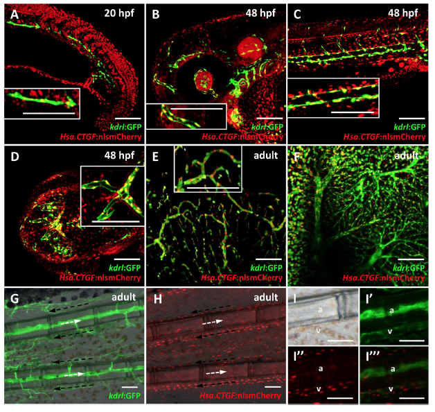
Fig. 4
Tg(Hsa.CTGF:nlsmCherry)ia49 reporter activity is prominent in the endothelium. (A–D) Reporter expression in the endothelium during development from 20 to 48 hpf. (E–I) Reporter expression in the adult endothelium. (A) Confocal sagittal section of the trunk of a 20 hpf Tg(Hsa.CTGF:nlsmCherry)ia49/Tg(kdrl:GFP) double transgenic embryo. Tg(kdrl:GFP) expresses GFP in all endothelial cells. Yap1/Taz are active in the endothelial cells in the developing vessels, as shown by the co-localization between the two signals. (B–D) Confocal Z-stack projections of the head region (side view in (B), dorsal view in (D)) and the trunk (C) at 48 hpf. Reporter signal co-localizes with kdrl:GFP expression throughout the embryo. (E,F) Confocal Z-stack projection of brain (E) and liver (F) tissue of a double transgenic adult fish, displaying Yap1/Taz reporter activation in the endothelium of respectively the cerebral and hepatic vascular networks. The insets represent zoomed views highlighting the co-localization between Hsa.CTGF:nlsmCherry and kdrl:GFP. (G–I) Fluorescent microscope images of adult caudal fin in Tg(kdrl:GFP) (G) and Tg(Hsa.CTGF:nlsmCherry)ia49 (H). Lateral views, anterior to the left, dorsal to the top. Arterial (white arrows) and venous (black arrows) bloodstream is indicated. Yap1/Taz reporter activity is stronger in the veins running laterally to the bony fin rays with respect to arteries inside the bony rays, as highlighted in the single channels and merge magnifications (I–I”’). a: artery; v-vein. Scale bar: 100 μm.

