Fig. S4
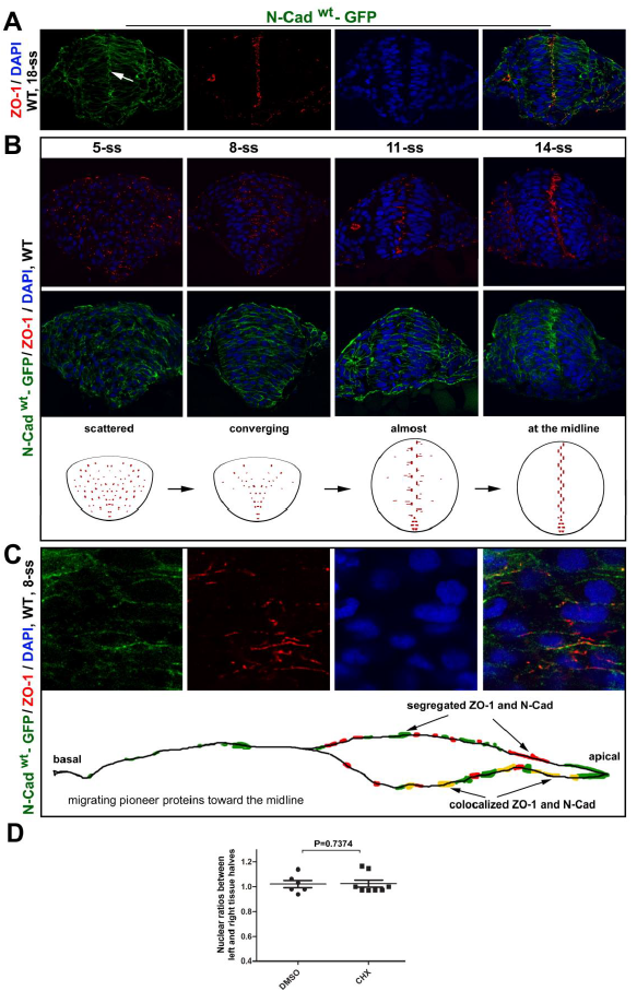
ZO-1 and N-Cadwt-GFP translocate apically during neural keel-neural rod transition in Tg(HSP70:N-Cadwt-GFP)pt137
A. Distributions of N-Cadwt-GFP and ZO-1 in the neural rod at 18-ss were revealed by immunohistochemistry of the transverse tissue sections. Note the apical enrichment of both proteins (arrows). N-Cadwt-GFP was briefly induced by a heat shock at 10-ss. B. Transverse imaging at 5-ss, 8-ss, 11-ss, and 14-ss: ZO-1 first localized to small punctate sites and then enriched at the midline region, whereas N-Cadwt-GFP distributed broadly on the cell membranes and then enriched at the midline region. Diagrams summarize the dynamics of ZO-1 distributions. C. A higher magnification to better reveal that ZO-1 foci, which were often closely associated with N-Cadwt-GFP, became more concentrated in the apical half of the cells at 8-ss and localized to larger and more elongated spots than at earlier stages. The bottom diagram depicts the distribution of ZO-1 and N-Cadwt-GFP on the cell membrane. D. Individual-value bar graphs (means ± SEM) show that the cycloheximide (CHX) treatment (see the protocol in Fig. 4E) did not affect the nuclear ratios between the left and right halves of the neural tissue. Numbers of embryos analyzed: 6 DMSO-treated; 8 CHX-treated. P values by two-tailed t test.

