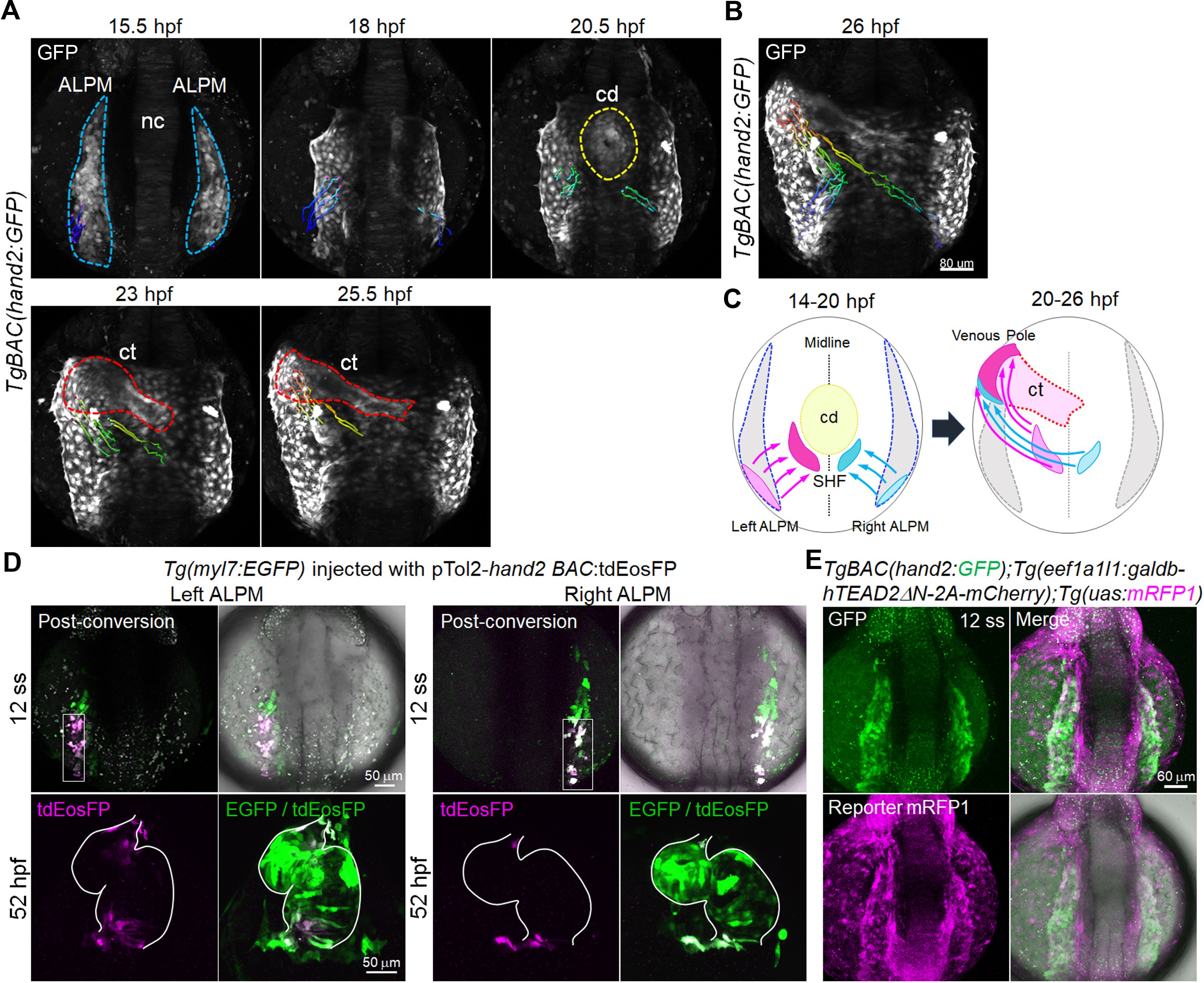
Fig. 4
Tead-reporter-active cells in the caudal region of the ALPM move to the venous pole.
(A) Time-sequential 3D-rendered confocal images of the TgBAC(hand2:GFP) embryo from 15.5 hpf to 25.5 hpf (n = 6). Spots of magenta and cyan denote the cells in the caudal part of the left and right ALPM, respectively. Notochord, nc; cardiac disc, cd; cardiac tube, ct. ALPM, cd, and ct are indicated by the blue, yellow, and red broken lines, respectively. (B) Tracking of caudal end hand2-promoter-active ALPM cells from 14 hpf to 26 hpf. The 3D-rendered confocal image of the TgBAC(hand2:GFP) embryo at 26 hpf with the track of cells showing color changes from blue to red according to the time after imaging (14 hpf to 26 hpf). (C) Schematic illustration of the trajectory patterns of the caudal end ALPM cells from 14 hpf to 26 hpf. Magenta and cyan denote the caudal region of the left and right ALPM, respectively. The cells in the caudal region of the left ALPM (magenta) and the right ALPM (cyan) moved to the venous pole (26 hpf) through the SHF region posterior to the cd (20 hpf). (D) Confocal 3D-stack images of the Tg(myl7:EGFP) embryo injected with pTol2-hand2 BAC:tdEosFP at the 12 somite stage (ss) (upper panels) and at 52 hpf (lower panels). Cells from the caudal region of either left ALPM (left four panels, n = 19) or right ALPM (right four panels, n = 18) were photoconverted at 12 ss. The hearts of the photoconverted embryos were imaged at 52 hpf. White squares and white lines indicate the photoconverted area and the outline of the heart, respectively. (E) Confocal 3D-stack images (at 12 ss) of the TgBAC(hand2:GFP);Tg(eef1a1l1:galdb-hTEAD2ΔN-2A-mCherry);Tg(uas:mRFP1) embryo (n = 6). Tead-reporter-active cells were present in the entire ALPM. Images of the ALPM are in dorsal view, anterior to the top. Images of the heart are in ventral view, anterior to the top. The confocal 3D-stack images are a set of representative images from at least five independent experiments.

