
|
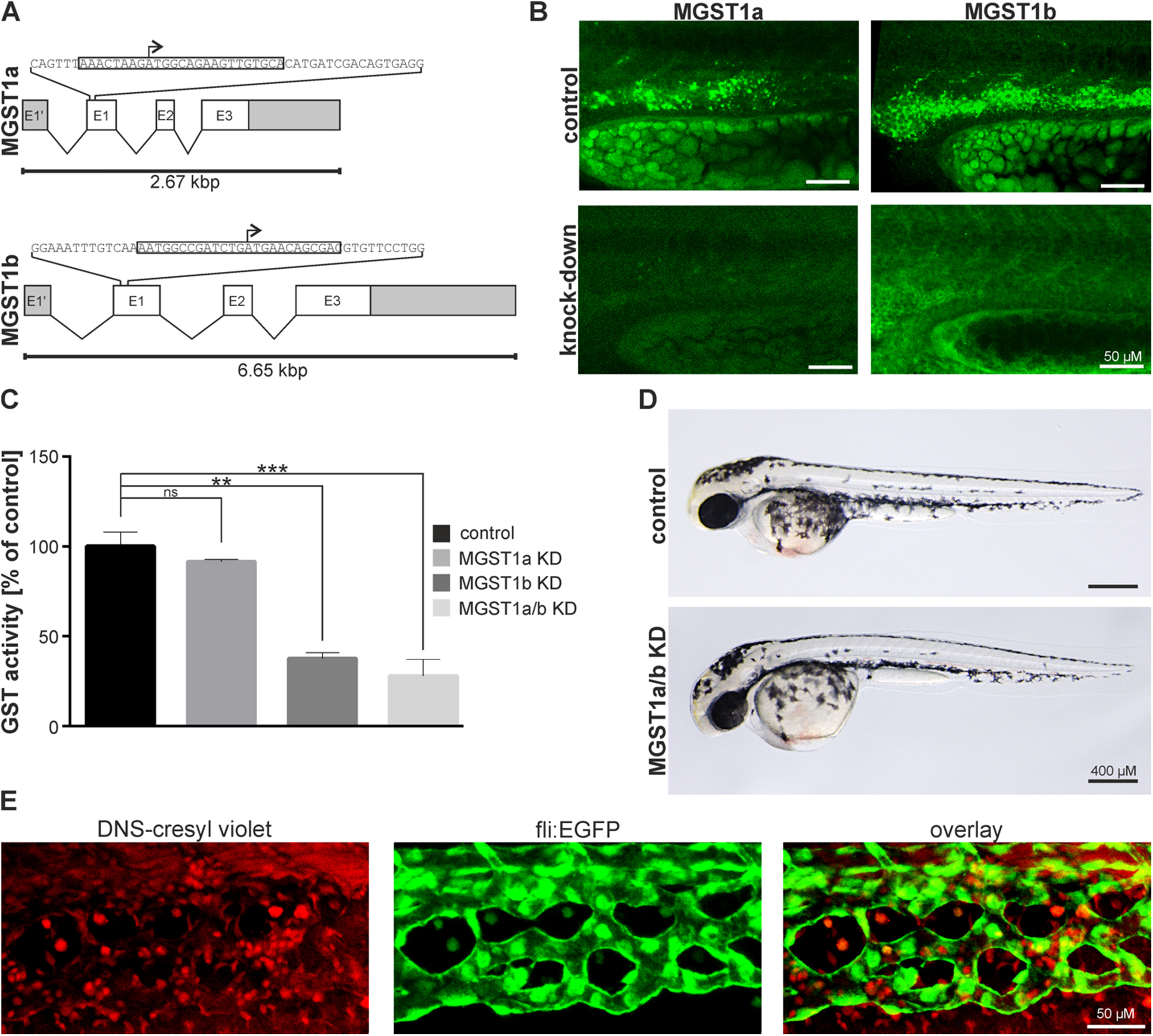
Fig. 2
Morpholino induced knock-down of MGST1 in zebrafish. (A) Genomic organization of zfMGST1 and location of morpholino attachment sites targeting transcription. (B) Immunohistochemistry in embryos injected with morpholinos knocking down MGST1a/b. (C) Enzymatic activity of GST enzymes in extracts of embryos injected with morpholinos knocking-down MGST1a, MGST1b or both. (D) Gross morphology of embryos injected with morpholinos knocking-down MGST1a/b. (E) DNS-cresyl violet staining indicating MGST1 positive cells in the caudal hematopoietic tissue, see also Supplementary movie S1. Brightfield images were taken on a Leica MZ16 microscope equipped with a Leica DFC300FX camera; Confocal images were taken on a Leica LSM700 with a 20× lens; Alexa 488 and 555 filters were used; images stacks were produced with ImageJ, GIMP was used to adjust the gamma channel.