
|
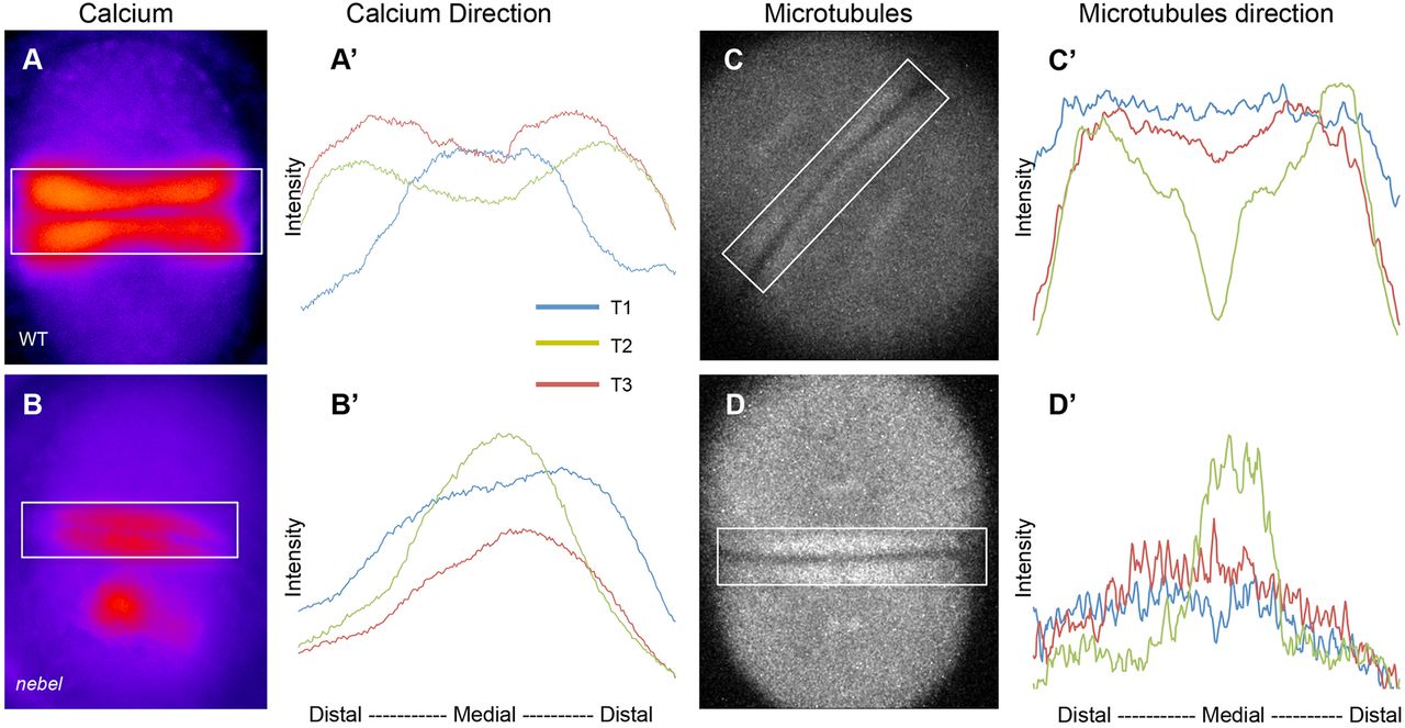
Fig. 7
FMA distal enrichment correlates with SCW directionality. (A-B′) Calcium levels in single Tg[βactin2:GCaMP6s] transgenic embryos during furrow progression, with plotted profiles (A′,B′) corresponding to boxed areas. Wild-type embryos (A′), but not nebel mutants (B′), show increasing distal calcium levels. (C-D′) Microtubules in EMTB::EGFP transgenic embryos during furrow progression (C,D), with plotted profiles (C′,D′). Wild-type embryos (C′), but not nebel mutants (D′), show distal enrichment. Time points T1, T2 and T3 correspond, respectively, to frames within one-tenth of the total sequence of acquired images spanning a furrow formation cycle at the beginning, middle and end of that cycle. See Fig. S4 for additional profiles.