Fig. 3-S2
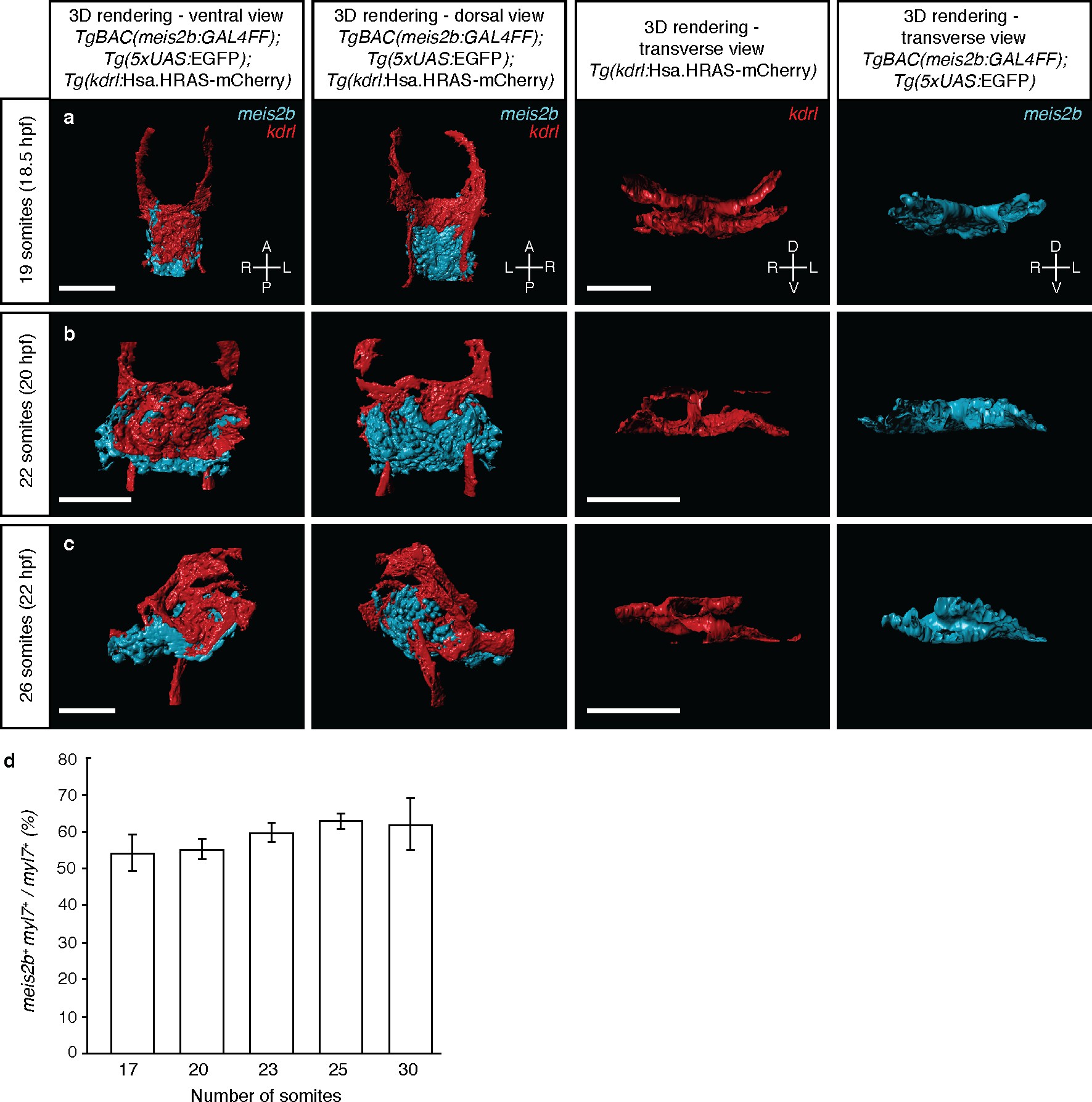
Tg(meis2b-reporter) expression with respect to the endothelium during the cardiac disc and heart tube stages.
3D surface rendering of Tg(meis2b-reporter);Tg(kdrl:Hsa.HRAS-mCherry) embryos at 19 to 26 ss. (a) At 19 ss, endocardial cells are located ventral to the myocardium and pass through the ring of myocardial cells to connect dorsally to the aortic arches. (b) At 22 ss, endocardial cells cover most of the ventral side of the myocardium in the cardiac disc. (c) At 26 ss, Tg(meis2b-reporter) expression is observed in the ventral side of the heart tube, while endocardial cells are lining the interior of the heart tube. Left column: ventral views, anterior up. Second column: dorsal views, anterior up. Third and fourth column: transverse views, dorsal up. (d) Percentage of Tg(meis2b-reporter)-positive cardiomyocytes with respect to the total number of myl7+ cardiomyocytes from 17 to 30 ss. Scale bars: 100 µm.

