Fig. 4
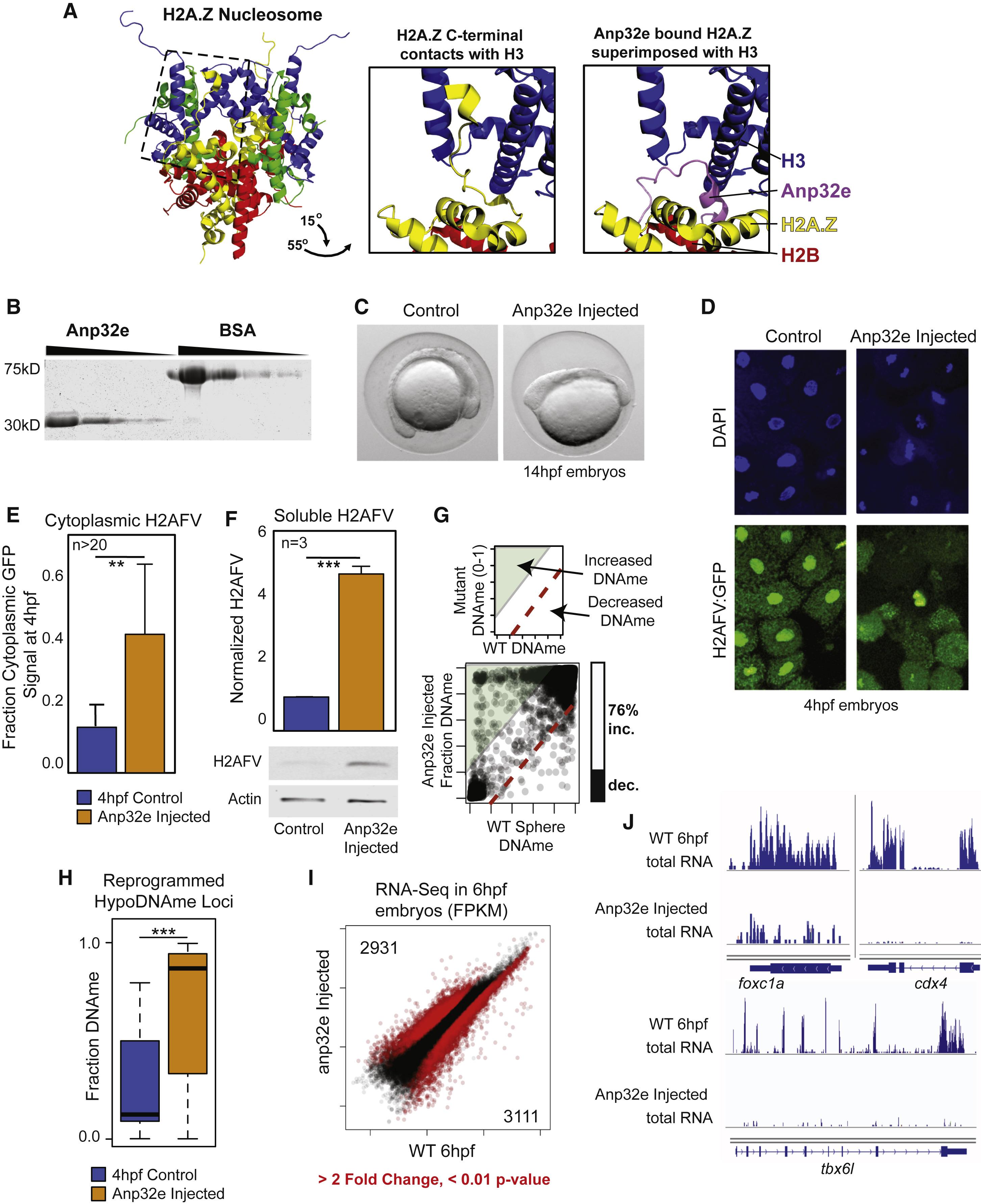
Anp32e Protein Injection Causes Reduced Nuclear H2AFV and Increased DNA Methylation
(A) Anp32e binding inhibits H2A.Z within the nucleosome. Left: crystal structure for the nucleosome octamer containing mouse H2A.Z (yellow) (Suto et al., 2000). Middle: zoomed-in and rotated view of the H2A.Z nucleosome octamer (dashed box from left), where the C terminus of H2A.Z interacts with histone H3. Right: the same view as middle panel, with the superimposed H2A.Z bound to human ANP32E (Obri et al., 2014).
(B) Purification of recombinant zebrafish Anp32e protein. A comparison to BSA and Coomassie staining to demonstrate purity.
(C) Phenotype of early somite-staged zebrafish following Anp32e protein injection.
(D) H2AFV signal is depleted in nuclei following Anp32e injection in embryos. DAPI and H2AFV:GFP localization in cells from sphere-staged (4hpf) embryos. Injection was into 1-cell embryos.
(E) Microscopy-based quantification of H2AFV following Anp32e injection. Fraction H2AFV:GFP signal in control and injected embryos like in (D). t tests: ∗∗ p < 0.01
(F) Quantification of H2AFV in cellular fractions resulting from Anp32e injection. Western blotting for soluble H2AFV indicates that Anp32e injection causes increased cytoplasmic H2AFV. t tests: ∗∗∗ p < 0.001.
(E and F) Error bars represent SD.
(G) Anp32e injection causes increased DNAme. Scatterplot of CpGs for treatment versus control is displayed, with a bar graph quantifying changing CpGs (>20% absolute change).
(H) Changes in DNA methylation are most significant at regions at which DNA is normally demethylated. t tests: ∗∗∗ p < 0.001. Boxplots are displayed.
(I) A scatterplot of gene transcript abundance (fragments per kilobase of million mapped reads [FPKM]) for Anp32e-injected embryos compared to control samples. Red data points indicate fold change >2 and p < 0.01 false discovery rate (FDR). The number of genes changing is indicated in the top-left and bottom-right corners.
(J) A gallery of browser snapshots for total RNA at developmentally regulated genes.
See also Figure S4.
Reprinted from Cell, 172(5), Murphy, P.J., Wu, S.F., James, C.R., Wike, C.L., Cairns, B.R., Placeholder Nucleosomes Underlie Germline-to-Embryo DNA Methylation Reprogramming, 993-1006.e13, Copyright (2018) with permission from Elsevier. Full text @ Cell

