
|
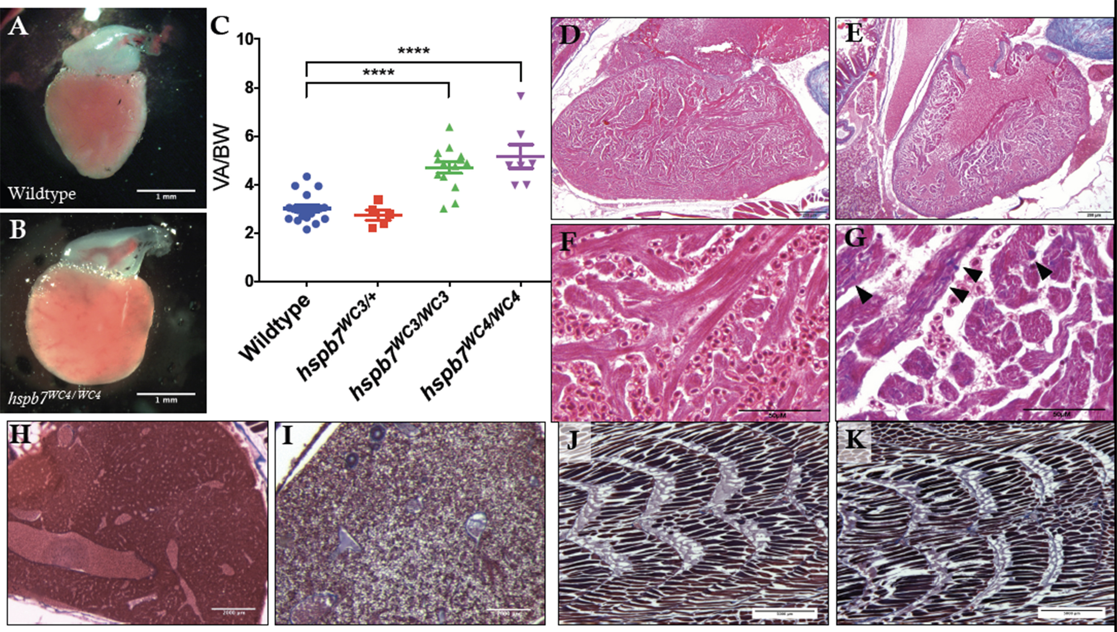
Fig. 4
Evidence of cardiomyopathy in hspb7WC3/WC3 adult hearts. Representative images of dissected ventricles and outflow tracts from (A) wildtype and (B) hspb7WC3/WC3 adults. Shown are hearts from 10 month old animals with the atria removed for clear viewing. (C) Quantification of ventricular area to body weight index (VA/BW) showed heart enlargement in animals homozygous for either of the hspb7 mutations. Each point represents one animal. Data are combined for 10 and 18 month zebrafish. Changes in heart size are significant according to an unpaired t-test, corrected for multiple comparisons with the Holm-Sidak method (p<0.0001). (D) Trichrome staining of 3 month wildtype and (E) hspb7 mutant hearts. Focal fibrotic lesions (arrowheads) were seen in all (5/5) hspb7 mutants but never in wildtype samples. Shown are representative images; F and G are higher magnification images for wildtype and mutant hearts, respectively. Arrowheads indicate local fibrotic lesions. (H-K) Representative Trichrome-stained images of adult wildtype (H,J) and mutant (I,K) hspb7WC3/WC3 zebrafish liver (H,I) and skeletal muscle (J,K).
Reprinted from Developmental Biology, 435(1), Mercer, E.J., Lin, Y.F., Cohen-Gould, L., Evans, T., Hspb7 is a Cardioprotective Chaperone Facilitating Sarcomeric Proteostasis, 41-55, Copyright (2018) with permission from Elsevier. Full text @ Dev. Biol.