
|
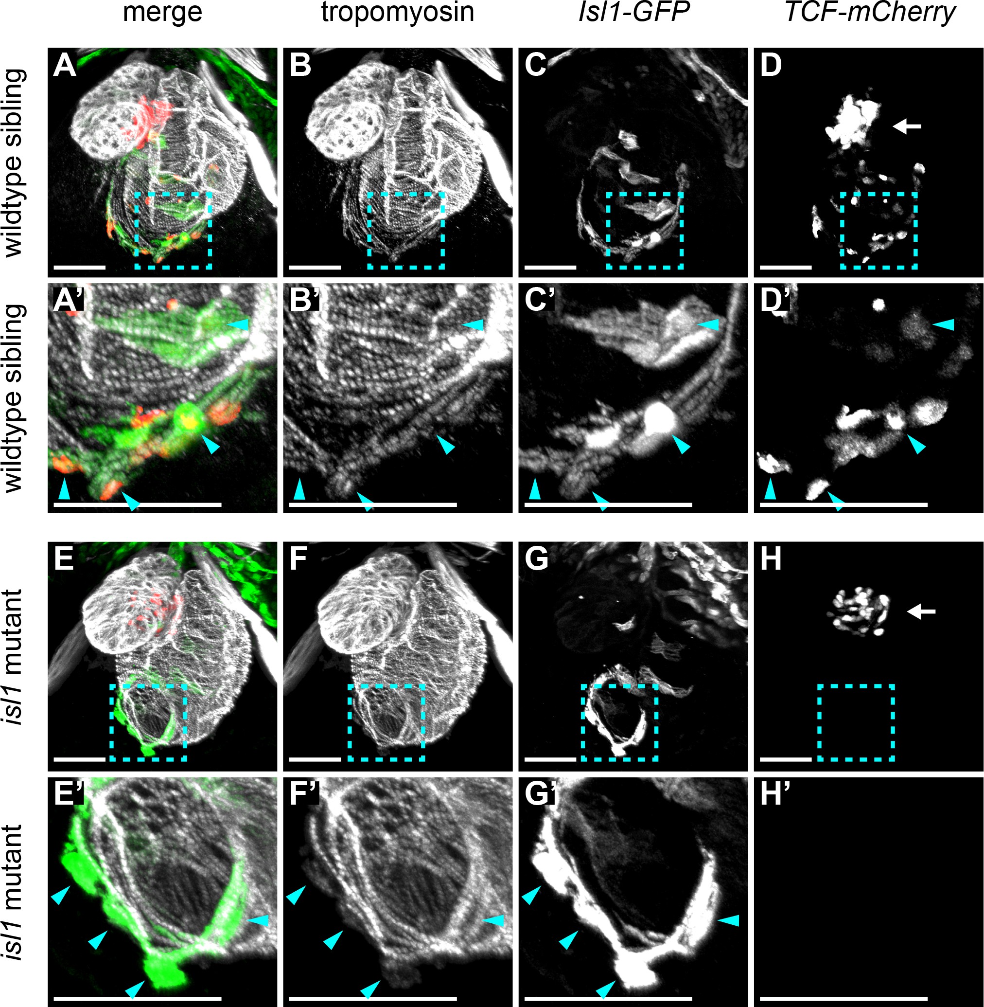
Fig. 5
Wnt/β-catenin signaling in cardiac pacemaker cells.
3D reconstructions of confocal scans from whole mount embryos containing the tg(7xTCFXla.Siam:nlsmCherry) (reporting Wnt/β-catenin activity in red) and the tg(Isl1:GFF;UAS:GFP) (reporting isl1 expression and marking pacemaker cells in green) and stained for tropomyosin (myocardium in white). Anterior is up and posterior is down. Wild-type sibling (A-D’) and isl1K88X mutant (E-H’) embryos at 3 dpf. The blue dashed line boxes in A-D and E-H indicate the sinoatrial region shown enlarged in A’-D’ and E’-H’, respectively. In wild-type hearts, Isl+ pacemaker cells (blue arrowheads) co-expressed TCF-mCherry. In isl1-/- mutants, no TCF-mCherry expression was observed in the Isl1+ pacemaker cells (blue arrowheads). TCF-mCherry expression was also detected in the AV canal of wild type (D) and isl1-/- mutants (H) (arrows). Scale bars represent 50 μm. See also Figure 5—figure supplement 1.