Image
Figure Caption
Fig. 4
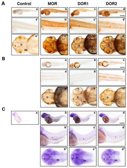
Antibodies against MOR, DOR1 and DOR2 recognize endogenous receptors in zebrafish embryos by immunohistochemistry. (A) Lateral and dorsal views of opioid receptors protein distribution in zebrafish embryos by whole-mount immunohistochemistry at 48 hpf. Embryos were processed as described in the Material and Methods section and subjected to immunohistochemistry with rabbit IgGs (Control), MOR, DOR1 or DOR2 antibodies for panels a, b, c and d, respectively. Magnified trunk (a′, b′, c′ and d′) and head panels (a′′, b′′, c′′ and d′′) are shown. Scale bar: 250 µm. Abbreviations: T: telencephalon; D: diencephalon; Hb: hindbrain; red asterisk: somite. A representative experiment is shown (n = 3); (B) Lateral and dorsal views of opioid receptors protein distribution in zebrafish embryos injected with the corresponding morpholino by whole-mount immunohistochemistry at 48 hpf. Embryos were injected with morpholinos specific to different opioid receptors at the one-to-four-cell stage in the yolk (panels a, b and c). Magnified trunk (a′, b′ and c′) and head panels (a′′, b′′ and c′′) are shown. Scale bar: 250 µm. Note the reduced signal in embryos injected with the corresponding morpholino compared to non-injected embryos (panel (A)). A representative experiment is shown (n = 3). (C) Lateral and dorsal views of opioid receptors mRNA distribution in zebrafish embryos by whole-mount in situ hybridization at 48 hpf. Expression of opioid receptor mRNA using no probe (a) and antisense probes against MOR (b), DOR1 (c) and DOR2 (d) are shown. Magnified trunk (b′, c′ and d′) and head panels (b′′, c′′ and d′′) are shown; red asterisk: somite. Scale bar: 250 µm. A representative experiment is shown (n = 3).
Figure Data
Acknowledgments
This image is the copyrighted work of the attributed author or publisher, and
ZFIN has permission only to display this image to its users.
Additional permissions should be obtained from the applicable author or publisher of the image.
Full text @ Int. J. Mol. Sci.

