Fig. 5-S1
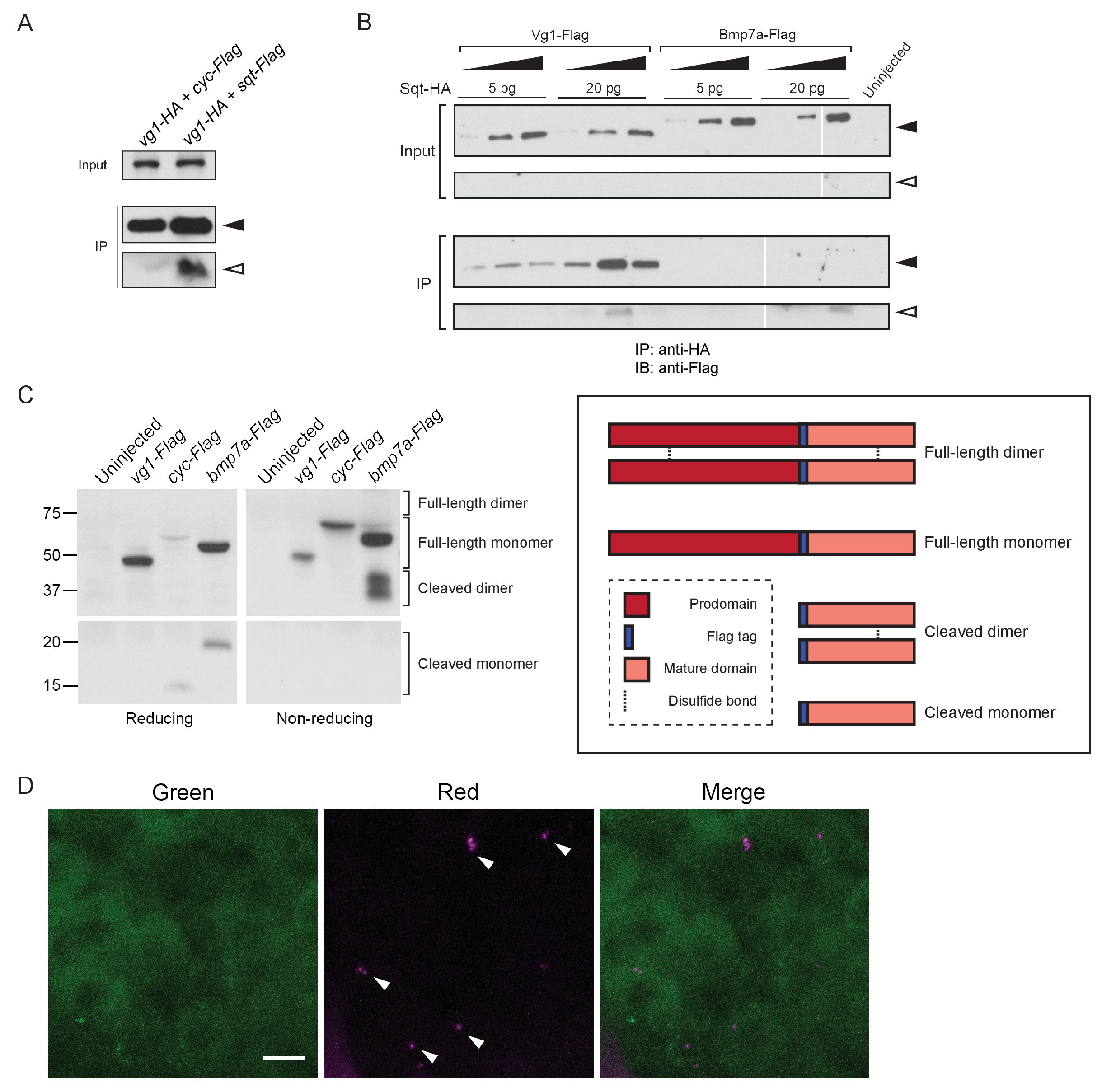
Vg1 and Nodal form heterodimers.
(A) Anti-Flag reducing immunoblot of anti-HA IP from lysates of WT embryos injected with 50 pg of vg1-HA mRNA and 50 pg of cyc-Flag or sqt-Flag mRNA. Black arrowhead indicates full-length protein; open arrowhead indicates cleaved protein. (B) Anti-Flag reducing immunoblot of anti-HA IP from lysates of Mvg1 embryos injected with either 5 pg or 20 pg of sqt-HA mRNA combined with increasing concentrations (5 pg, 20 pg or 50 pg) of vg1-Flag or bmp7a-Flag mRNA. Black arrowhead indicates full-length protein; open arrowhead indicates cleaved protein. In contrast to Vg1-Flag, the mature domain of Bmp7a-Flag is more readily detected than the full-length protein (Little and Mullins, 2009). The samples were loaded onto three gels run under the same conditions at the same time (gel boundaries are marked by white vertical lines). (C) Anti-Flag reducing and non-reducing immunoblots of Mvg1 embryos injected with vg1-Flag, cyc-Flag or bmp7a-Flag mRNA (from Figure 5B). Approximate locations of expected monomers and dimers are indicated. TGF-beta family protein dimers are connected by disulfide bonds, which are broken under reducing conditions. (D) Z-stack of ~256-cell stage Mvg1 embryo co-injected with 100 pg of vg1-Dendra2 mRNA at the 1-cell stage and 5 pg of cyc mRNA at the 64-cell stage immediately followed by photoconversion. The red puncta represent Vg1-Dendra2 protein synthesized before the 64-cell stage that has heterodimerized and been secreted with Cyc protein.

