
|
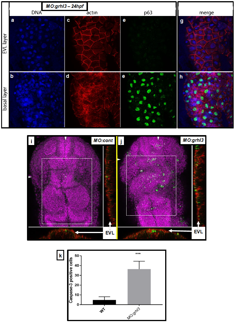
Fig. S3
Characterisation of cell identity and viability in the EVL and basal layers of MO:grhl3 injected fish. (A-H) DAPI staining showing no loss of nuclei in either EVL or basal layers (A-B). Rhodamine-Phalloidin staining showing normal distribution of F-actin in the EVL (C) and basal layer (D). There are no ectopic basal keratinocytes (p63+) present in the EVL (E), although, as expected, these are abundant in the basal layer (F; merged views G-H). (I-K) Activated Caspase-3 immunohistochemistry reveals few apoptotic cells in control fish (I) relative to the significantly increased numbers of apoptotic cells seen in MO:grhl3 injected embryos (J) at the level of the midbrain and hindbrain (boxed region in I-J); orthogonal views at the levels indicated (arrowheads; I-J) confirm that the majority of apoptotic cells are seen in the EVL and adjoining dorsal-most neural tissue, but are largely absent from the deeper neural tissue. These data are quantitated from n=4 controls and n=3 MO:grhl3-injected embryos (K).