
|
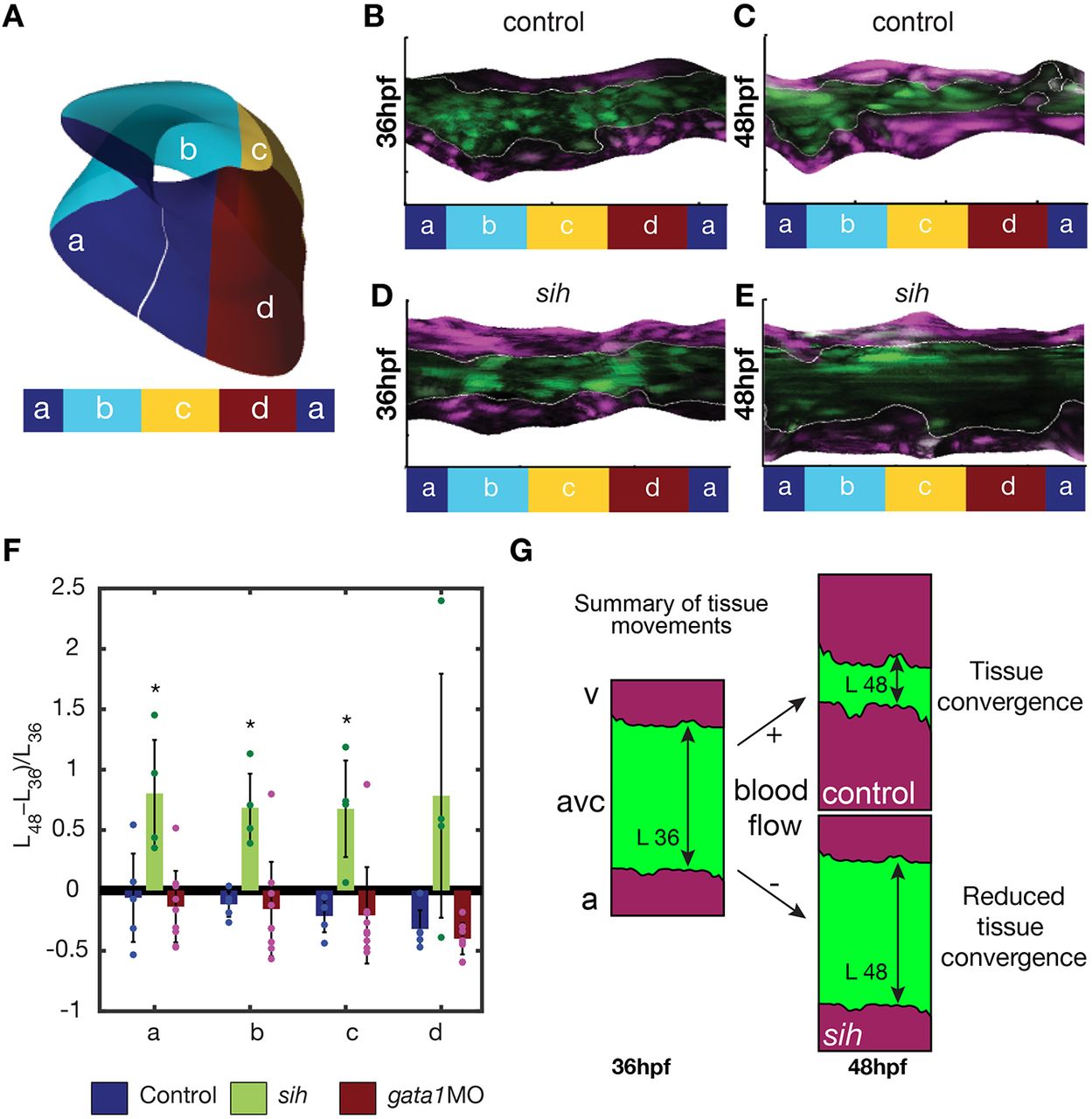
Fig. 2
Tissue convergence (AVC shortening) is flow dependent. (A) Schematic showing the compartmentalization into superior (a), inferior (c), exterior (b) and interior (d) regions of the AVC. (B-E) Unfolded visualization of the fli:kaede (control) (B,C) and sih;fli:kaede AVC endocardium (D,E) at 36 hpf (B,D) and 48 hpf (C,E). (F) The shortening index (L48−L36)/L36 of the AVC between 36 hpf and 48 hpf in control (n=5), sih (n=4) and gata1 (n=9) morphants is averaged on the a,b,c,d regions. L48 and L36 are the AVC length L at 48 hpf and 36 hpf, respectively. Data are mean±s.d. Statistical significance was determined by unpaired Student's t-test; *0.01<P<0.05. (G) Schematic of flow-dependent tissue convergence.