
|
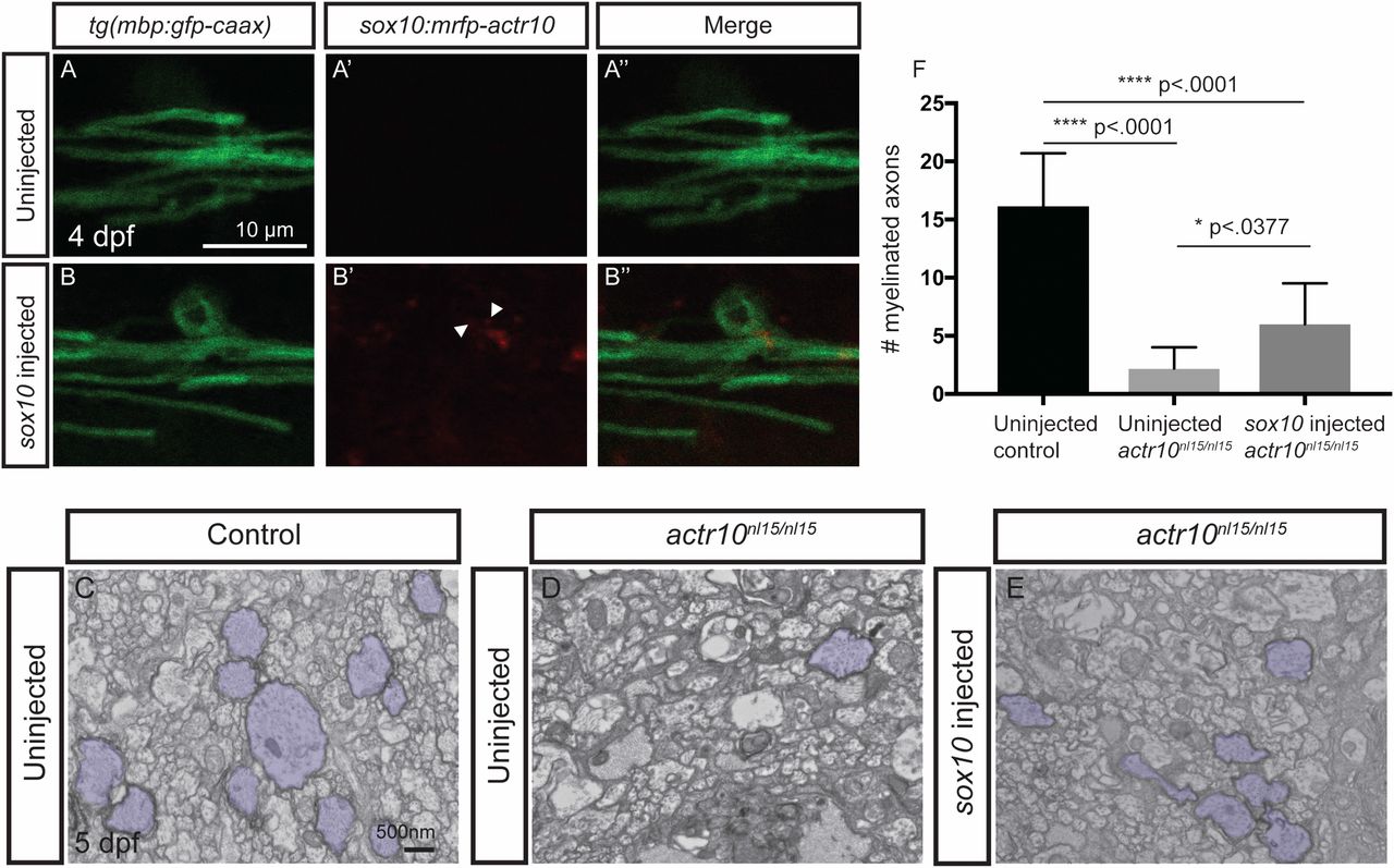
Fig. 5
Transient expression of actr10 in oligodendrocytes partially suppresses myelination defects in actr10nl15/nl15 mutants. (A–B′′) Confocal images of mbp-labeled oligodendrocytes in uninjected (A) and sox10:mRFP-actr10 (“sox10 injected”) actr10nl15/nl15 mutants (B). While uninjected actr10nl15/nl15 mutants do not exhibit RFP fluorescence (A′ and A′′), injection of sox10:mRFP-actr10 results in monomeric RFP fluorescence in actr10nl15/nl15 mutants (B′ and B′′). (C–E) TEM images show dorsal spinal cords of uninjected WT and actr10nl15/+ controls (C), uninjected actr10nl15/nl15 mutants (D), and sox10:mRFP-actr10–injected actr10nl15/nl15 mutants (E). Myelinated axons are pseudocolored in purple. (F) Quantification shows that sox10-injected actr10nl15/nl15 mutants (n = 6) have significantly greater numbers of myelinated axons in the dorsal spinal cord compared with uninjected actr10nl15/nl15 mutant siblings (n = 5, P < 0.0377), although sox10:mRFP-actr10 injection does not restore myelination to WT control levels (n = 5, P < 0.0001), indicative of partial rescue. One-way ANOVA with Tukey’s multiple comparisons test was used for statistical analyses.