Fig. 2
Fig. 2
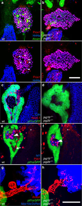
Jag1b and Jag2b are required for intrahepatopancreatic duct lineage specification. a–h Whole organ immunofluorescence expression analysis of the 72 hpf foregut endoderm of wild type and jag1b −/− ;jag2b −/− mutant embryos. (a, b Alcam channel removed in bottom panels). Intrahepatic duct cells marked by high Alcama levels (a, green), and by Prox1 (red) but not Hnf4a expression (blue, a), are found in wild type liver (arrowheads, a). These duct cells are not found in the jag1b −/− ;jag2b −/− mutant liver (b, representative sample, n = 13), which is comprised entirely of hepatocytes expressing both Prox1 and Hnf4a. c–f Intrapancreatic ductal cells expressing Nkx6.1 (red, c, d) or Pdx1 (red, arrow, e, f) are present in wild type but not in jag1b −/− ;jag2b −/− mutants. Pdx1 expression in Islet1+ cells (blue, arrowheads) and the duodenum (asterisk) is comparable to wild-type siblings, as is expression of ptf1a:GFP+ (green) acinar cells c–f and Anxa4+ (red) extrahepatopancreatic duct cells (red, g, h). g, h Anxa4+ (red) extrahepatopancreatic ducts can be found joining the ptf1a:GFP+ (green) pancreatic acinar cells to the fabp10a:DsRed+ hepatocytes (blue). Representative samples from three different jag1b −/+ ;jag2b −/+ in-cross clutches. Scale bars, 50 μM

