
Image
|
Figure Caption
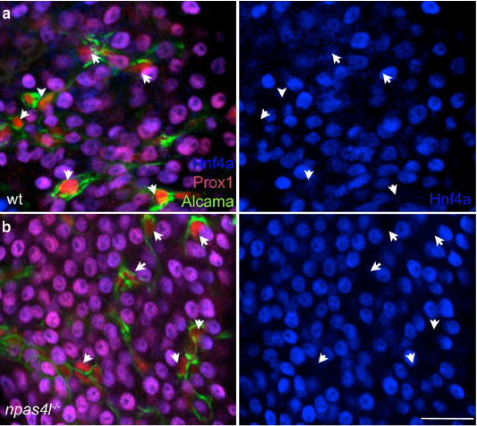
Fig. s6 Supplementary figure 6. Specification of intrahepatic duct lineage in the absence of the vasculature. (a-b) Whole organ immunofluorescent analysis of 76 hpf wild type (a) and npas4l mutant (b) livers labelled for Prox1 (red), Hnf4a (blue), and Alcama (green). Green Alcama channel is removed in panels below a and b. Intrahepatic duct cells (arrows), expressing both Prox1 and high Alcama, but lacking Hnf4a, are present in both wild type and mutant livers. Scale bars 20μM
Acknowledgments
This image is the copyrighted work of the attributed author or publisher, and
ZFIN has permission only to display this image to its users.
Additional permissions should be obtained from the applicable author or publisher of the image.
Full text @ Nat. Commun.