
|
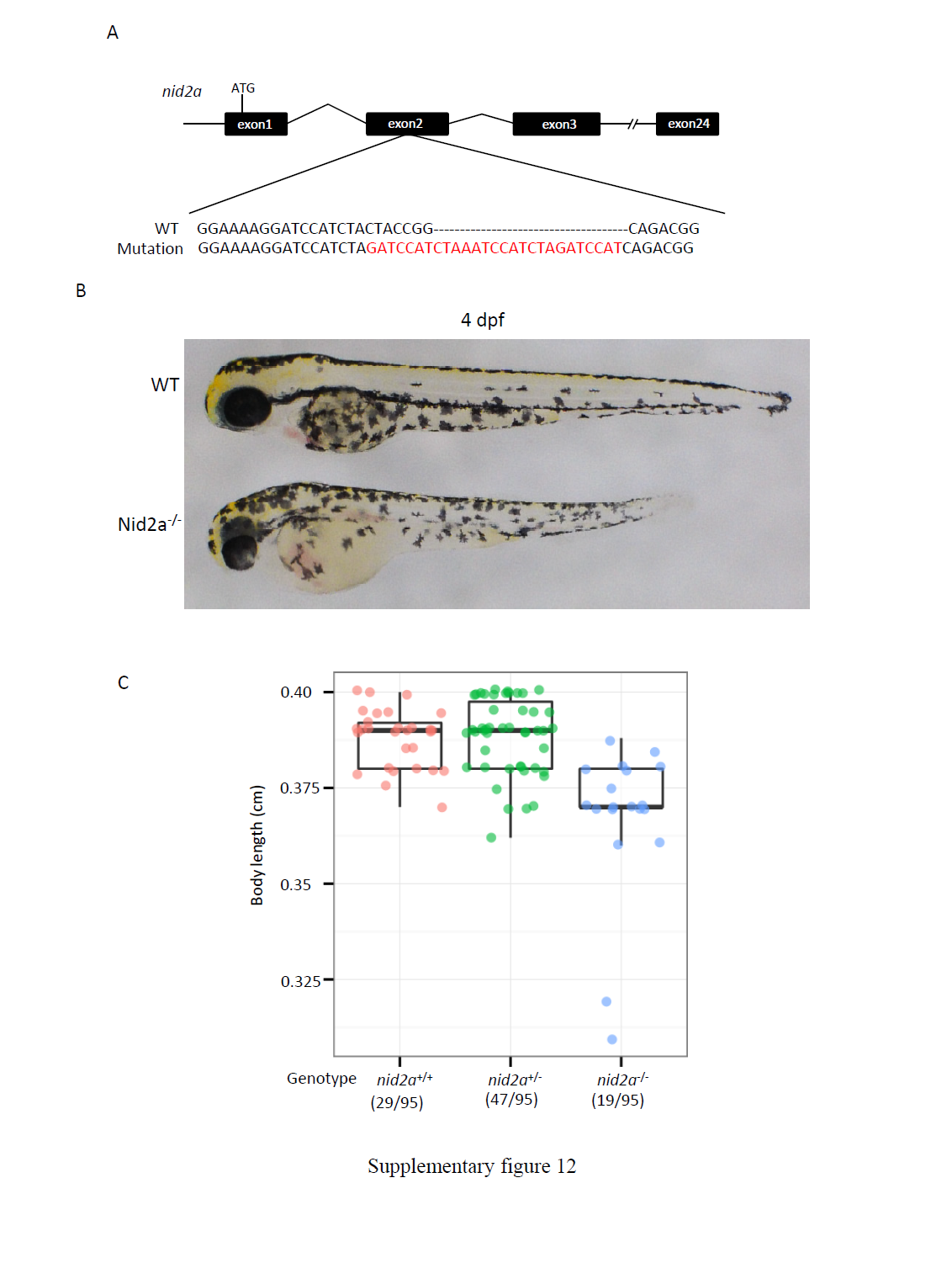
Fig. S12
nid2a knockout mutants display similar phenotypes to those in pIDM-A1 embryos upon Dox treatment. (A) Upper panel: Diagram showing the genome structure of nid2a and the gRNA target site. Bottom panel: Comparison of genomic DNA among WT and nid2a-/- mutant (with a 7 bp-deletion and a 27 bp-insertion in the 2nd exon ). ATG: translation start codon. The mutation will lead to an early stop codon. (B) Picture of WT and nid2a homozygous mutant (nid2a-/-) embryos at 4 dpf. (C) Statistics of body length (centimeter cm) of different genotype embryos in a F22 population at 4 dpf as indicated. Numbers: embryos with the genotype versus total embryos in the F22 population examined.