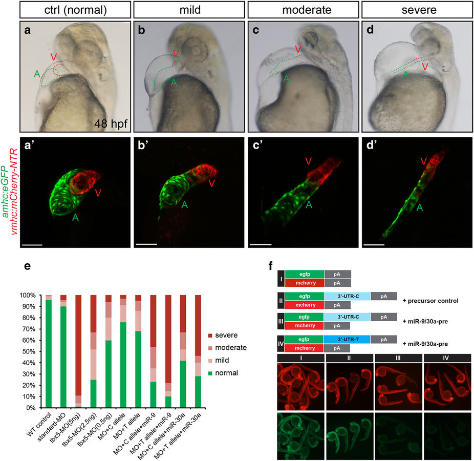
Fig. 4
TBX5 3′UTR variant rs6489956 affected heart development in zebrafish embryo. (a–d′) Examples of heart defects with variable severity in zebrafish: (a and a′) normal heart; (b and b′) mild defect: heart shows mild looping arrested, pericardium effusion, and dropsy of ventral position; (c and c′) moderate defect: heart shows moderate looping arrested, pericardium effusion and dropsy of ventral position; (d and d′) severe defect: malformed embryo with small, string-like heart, severe pericardium effusion, and dropsy of ventral position. Green A indicates ventricle; red V indicates atrium. (e) Distribution of four categories of heart morphologies in each experimental group at 48 hpf: WT control, MO standard control, MO (5 ng), MO (2.5 ng), MO (0.5 ng), MO+C allele mRNA, MO+T allele mRNA, MO+C allele mRNA+miR-9, MO+T allele mRNA+miR-9, MO+C allele mRNA+miR-30a, MO+T allele mRNA+miR-30a. (f) EGFP sensors were coinjected with mCherry control as indicated. miR-9/30a precursor injection reduced EGFP levels in EGFP-3′UTR sensor (third column), while mCherry levels were unchanged. In the EGFP-3′UTR-T sensor (fourth column), more significant reduction in EGFP was noted.

