Fig. 4
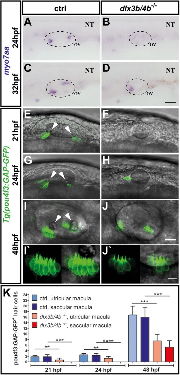
Delayed formation of sensory hair cells in dlx3b/4b-deficient embryos. (A-D) In comparison with control embryos at 24 hpf and 32 hpf, myo7aa expression is initially absent and subsequently restricted to the domain of the future utricular macula in dlx3b/4b mutants. (E-J) Expression of Tg(pou4f3:GAP-GFP) in control siblings and dlx3b/4b mutants at 21 hpf, 24 hpf and 48 hpf confirms delayed formation of sensory hair cells in dlx3b/4b-deficient embryos. Arrowheads indicate positions of the nascent otoliths in wild-type embryos. Note the initial onset of GFP in the prospective domain of the utricular macula and the subsequent expression in the future domain of the saccular macula in dlx3b/4b mutants. (I′,J′) Kinocilia formation appears normally in control siblings and dlx3b/4b mutants. Lateral views are shown with anterior to the left. NT, neural tube; OV, otic vesicle. Scale bars: 40 µm in D; 25 µm in J; 10 µm in J′. (K) Time course showing the mean number of pou4f3:GAP-GFP-positive hair cells in the domain of the future utricular and saccular domain of control (ctrl) and dlx3b/4b mutant embryos. dlx3b/4b-deficient embryos exhibit a significantly reduced number of sensory hair cells at 21 hpf and 24 hpf, and display significantly fewer hair cells at 48 hpf (**P<0,01; ***P<0,001; ****P<0,0001). Data are mean±s.e.m (n≥6 for each time point).

