Fig. 2
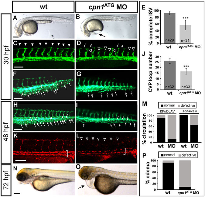
Knockdown of cpn1 causes vascular defects in zebrafish development. (A,B) The bright field images of control and cpn1 ATG morpholino (3.41 ng)-injected embryos at 30 hpf. (C–J) At 30 hpf, ISVs reached DLAV and formed a honeycomb-like structure in the caudal vein plexus (CVP) in wild-type controls by using Tg (fli1:eGFP y1) zebrafish (C,F). Compared with wild-type control (arrowheads in C, arrows in F), loss of cpn1 caused ISV growth defects (hollow arrowheads in D) and less honeycomb-like structure formation in CVP (arrows in G). At 48 hpf, less CVP loop formation (arrows in I) compared with that in control (arrows in H). (K,L) The injection of cpn1 ATG MO into Tg (fli1a:eGFP y1; gata1:dsRed sd2) embryos revealed that loss of cpn1 caused circulation defects in ISVs at 48 hpf. (N,O) At 72 hpf, cpn1 ATG morphants had pericardial edema (arrow in O). (E) The percentage of completed ISVs decreased by approximately 45% in cpn1 ATG morphants (n = 29 in wt and n = 31 in cpn1 ATG MO) at 30 hpf. (J) The loop formation in CVP exhibited a decrease in cpn1 ATG morphants (n = 32 in wt and n = 33 in cpn1 ATG MO) at 48 hpf. (M) Quantification data showed cpn1 morphants had approximately 90% defects in the ISV–DLAV circulation and 40% defects in the aorta–vein circulation compared to controls (n = 39 in control and n = 28 in cpn1 MO) at 48 hpf. (P) The percentage of pericardial edema was 90% in cpn1 ATG morphants compared with that in controls (n = 127 in controls and n = 50 in cpn1 ATG MO) at 72 hpf. These results were confirmed by three independent experiments. Data are represented as means ± S.D. ***Refers to p < 0.0001 by an unpaired Student’s t-test. The scale bar is 200 µm for A, B, N, and O, and 100 µm for C–L.

