Fig. 1
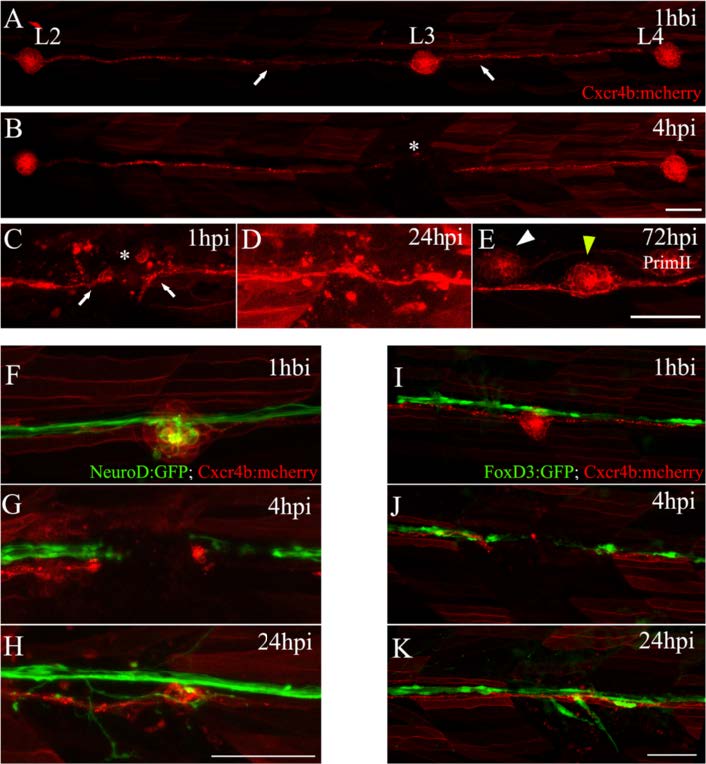
Electroablation as a method for localized tissue injury in the posterior lateral line (PLL) of zebrafish larvae. a Trunk of a tg(cxcr4b:mCherry) larva showing red-labeled PLL cells, including the second, third, and fourth neuromasts of the PLL (L2, L3, and L4) connected by interneuromastic cells (INCs, white arrows). The image was captured 1 hour before injury (hbi). b The trunk of the same larva 4 hours post injury (hpi). The asterisk shows the damaged zone, where the L3 neuromast was located. c–e Higher magnifications of the injured area showing the process of neuromast regeneration. c At 1 hpi, we observed the gap generated between INCs (white arrows) flanking the injury zone (asterisk). d At 24 hpi, INCs located on both sides of the gap reconnected. e At 72 hpi, the L3 neuromast had regenerated (yellow arrowhead). At this stage, the secondary primordium (PrimII) deposited a secondary neuromast (white arrowhead). f–h Double transgenic tg(neurod:GFP; cxcr4b:mCherry) larvae, where the afferent lateral line neurons are labeled in green and neuromasts are labeled in red. f This image shows the L3 region 1 hbi. g Electroablation of L3 interrupts the lateral line nerve. h The nerve regenerates after 24 hpi. i–k Double transgenic tg(foxd3:GFP; cxcr4b:mCherry) larvae, showing the Schwann cells labeled in green (associated with the nerve) and neuromasts and INCs labeled in red. As occurs with the INCs, Schwann cells reconnected after 24 hpi (k). Scale bar: 50 μm

