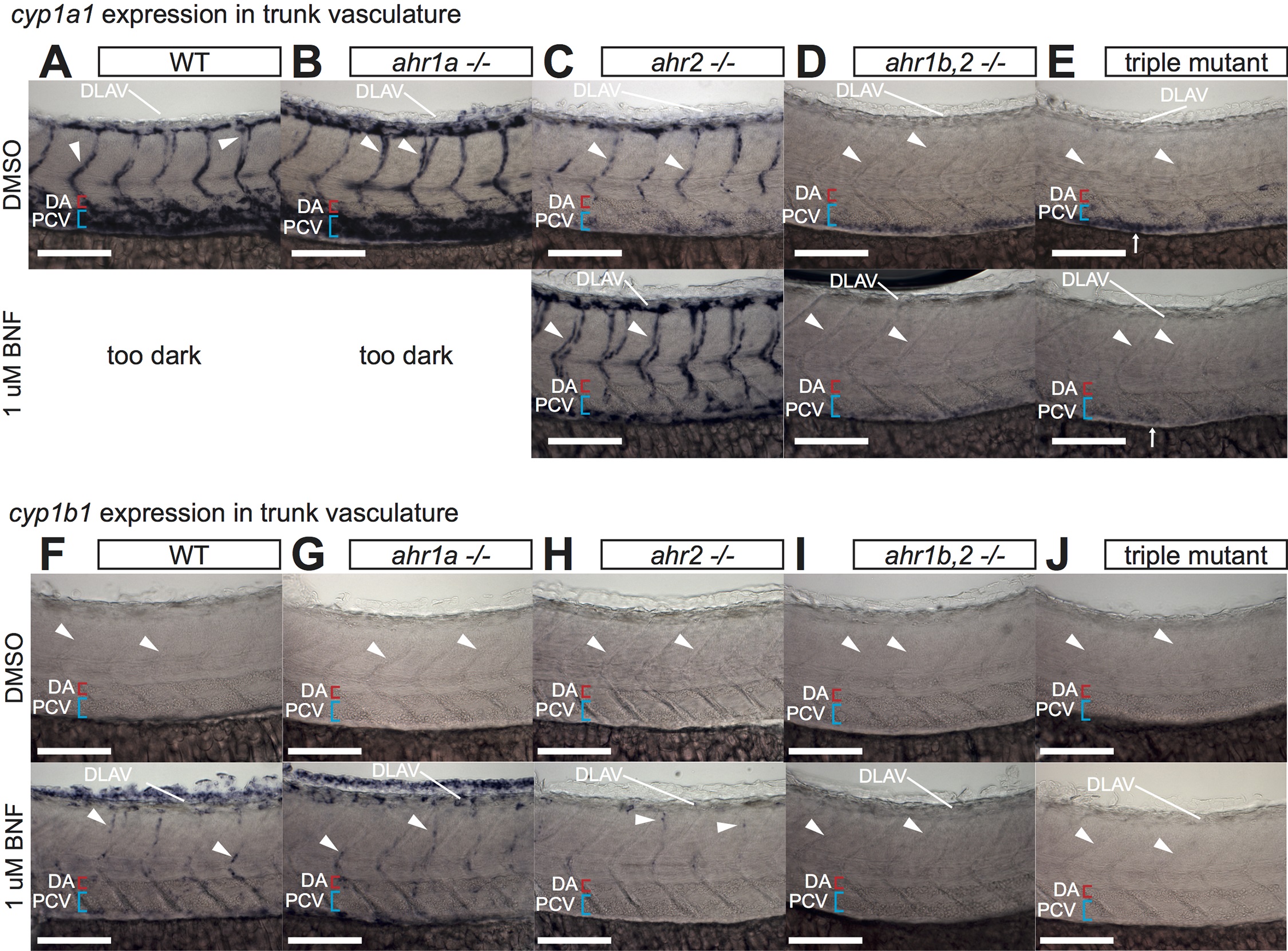
Fig. S3
Analysis of vascular CYP1 expression in trunk of AHR mutants.
A-J) High magnification images of cyp1a1/b1 expression in the trunk vasculature at 52 hpf in DMSO or BNF-treated WT (A, F), ahr1a -/- (B, G), ahr2 -/- (C, H), ahr1b,2 -/- (D, I) and triple AHR mutants (E, J). Expression patterns of both genes in ahr1a -/- are indistinguishable from WT in either condition. In ahr2 mutants a dramatic loss of endogenous cyp1a1 expression from the PCV and DA is observed, together with a weaker but persistent expression in DLAV and ISVs that is enhanced by BNF treatment (C). This remaining vascular expression is lost in ahr1b,2 -/- and triple AHR mutants (D, E). Note the AHR-independent expression of cyp1a1 in the gut (arrows in E) Similar results are seen in the BNF-induced cyp1b1 expression, which is weakly maintained in ISVs and DLAV of ahr2 mutants and lost in ahr1b,2 -/- and triple AHR mutant embryos (F-J). Numbers of embryos analyzed are the same as in Fig 4 (cyp1a1) and Fig 5 (cyp1b1). All scale bars are 100 um. Abbreviations–AHR: aryl hydrocarbon receptor, BNF: beta-naphthoflavone, CYP: cytochrome p450, DA: dorsal aorta, DLAV: dorsal longitudinal anastomotic vessel, DMSO: dimethylsulfoxide, hpf: hours post fertilization, ISV: intersegmental vessel, PCV: posterior cardinal vein, WT: wildtype.

