
|
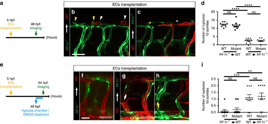
Fig. 5
hif-1α does not play a cell-autonomous role in ECs during DLAV plexus formation or during blood vessel repair after hypoxia.
(a) Schematic representation of the experiment shown in b–d. (b,c) Transplantation of hif-1α−/− Tg(kdrl:EGFP) donor cells into WT Tg(kdrl:ras-mCherry) host blastulae and of WT Tg(kdrl:ras-mCherry) donor cells into hif-1α−/− Tg(kdrl:EGFP) host blastulae shown at 48 hpf; white arrowheads point to sprouts from WT DLAV ECs, yellow arrowheads point to sprouts from hif-1α−/− DLAV ECs, white asterisk indicates missing sprout from a WT EC and yellow asterisk indicates missing sprout from a hif-1α−/− EC. (d) Quantification of DLAV sprouts in a 10 somite-long trunk area from WT and hif-1α−/− DLAV ECs in the two different EC transplantation conditions at 48 hpf. (e) Schematic representation of the experiment shown in f–i. (f–h) Transplantation of hif-1α−/− Tg(kdrl:EGFP) donor cells into WT Tg(kdrl:ras-mCherry) host blastulae, and of WT Tg(kdrl:ras-mCherry) donor cells into hif-1α−/− Tg(kdrl:EGFP) host blastulae shown after hypoxia chamber or DMOG treatment for 6 h starting at 48 hpf; white arrow points to vessel rupture affecting WT EC, and yellow arrow points to vessel rupture affecting hif-1α−/− EC. (i) Quantification of blood vessel ruptures in a 10 somite-long trunk area affecting WT and hif-1α−/− ECs in the two different EC transplantation conditions at 54 hpf after hypoxia chamber or DMOG treatment for 6 h starting at 48 hpf. Bars represent mean±s.e.m., n=10 embryos from three different clutches, (**P<0.01; ***P<0.001; ****P<0.0001; NS, no significant changes observed; t-test). Scale bars, 50 μm.