
|
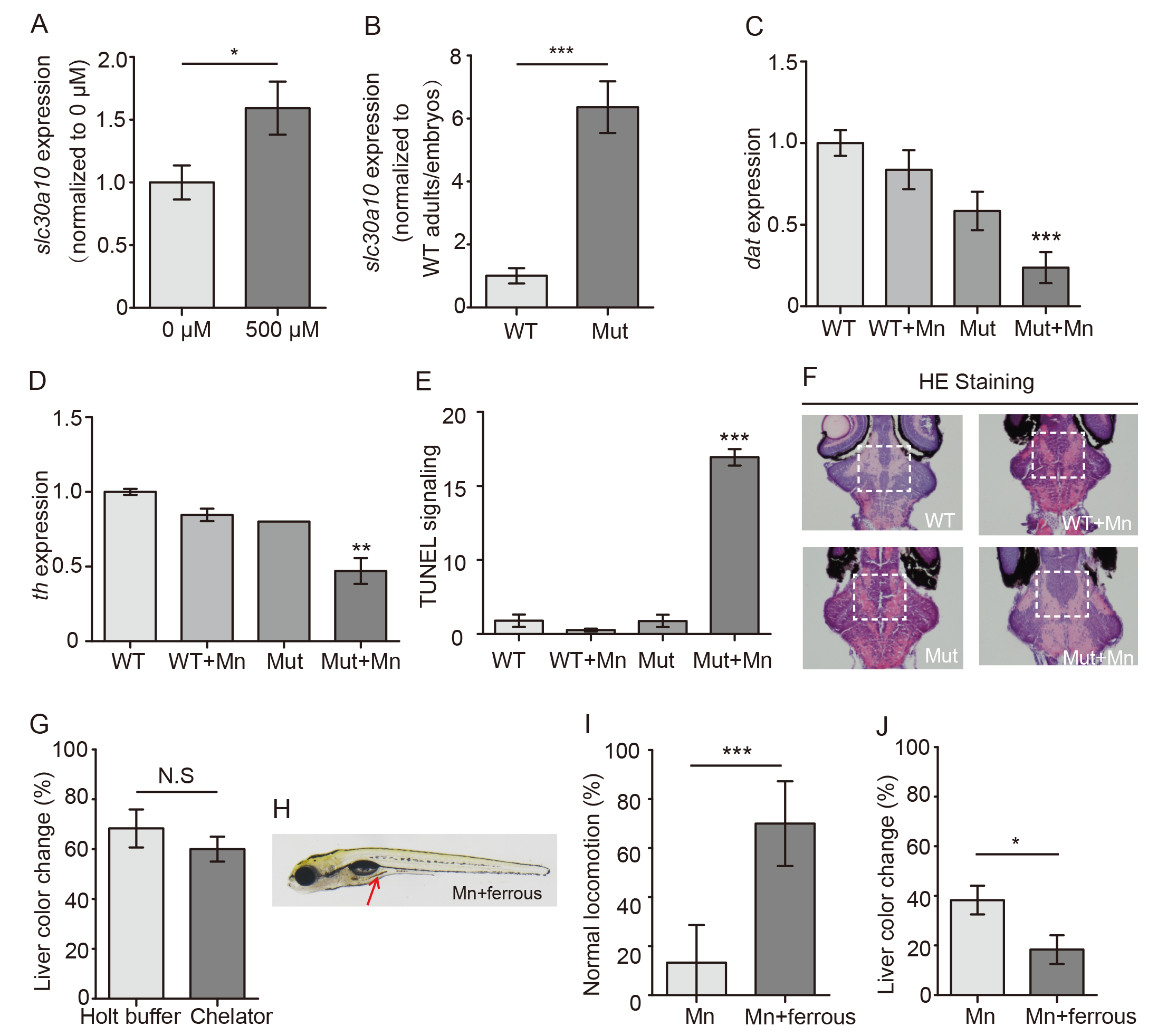
Fig. S3
Phenotypic characterization and effect of ferrous fumarate on slc30a10 mutant embryos.
(A) Slc30a10 mRNA was measured in heterozygous adults with or without Mn exposure. (B) Slc30a10 mRNA level was showed by a ratio of adults versus embryos in both wild-type and mutants. (C and D) Quantitative analyses of in situ staining for dat (C) and th (D) mRNA in WT and mutant embryos; where indicated, the embryos were exposed to Mn. (E) Quantitative analyses of TUNEL fluorescence. (F) HE staining in frozen sectioned brain of embryos. (G) Following Mn exposure, mutant embryos were transferred to either fresh Holt buffer of buffer containing the chelator EDTA-CaNa2, and the percentage of embryos with a color change in the liver is plotted (n = 3 sets of 20 embryos/group). (H) Image of a Mn-exposed mutant embryo after treatment with ferrous fumarate; note the brown color in the gut (arrow). (I and J) Summary of locomotion (I) and the percentage of embryos with a color change in the liver (J) in mutant Mn-exposed embryos treated with ferrous fumarate (n = 3 sets of 20 embryos/group). *p<0.05, **p<0.01, and ***p<0.001.