Image
Figure Caption
Fig. S1
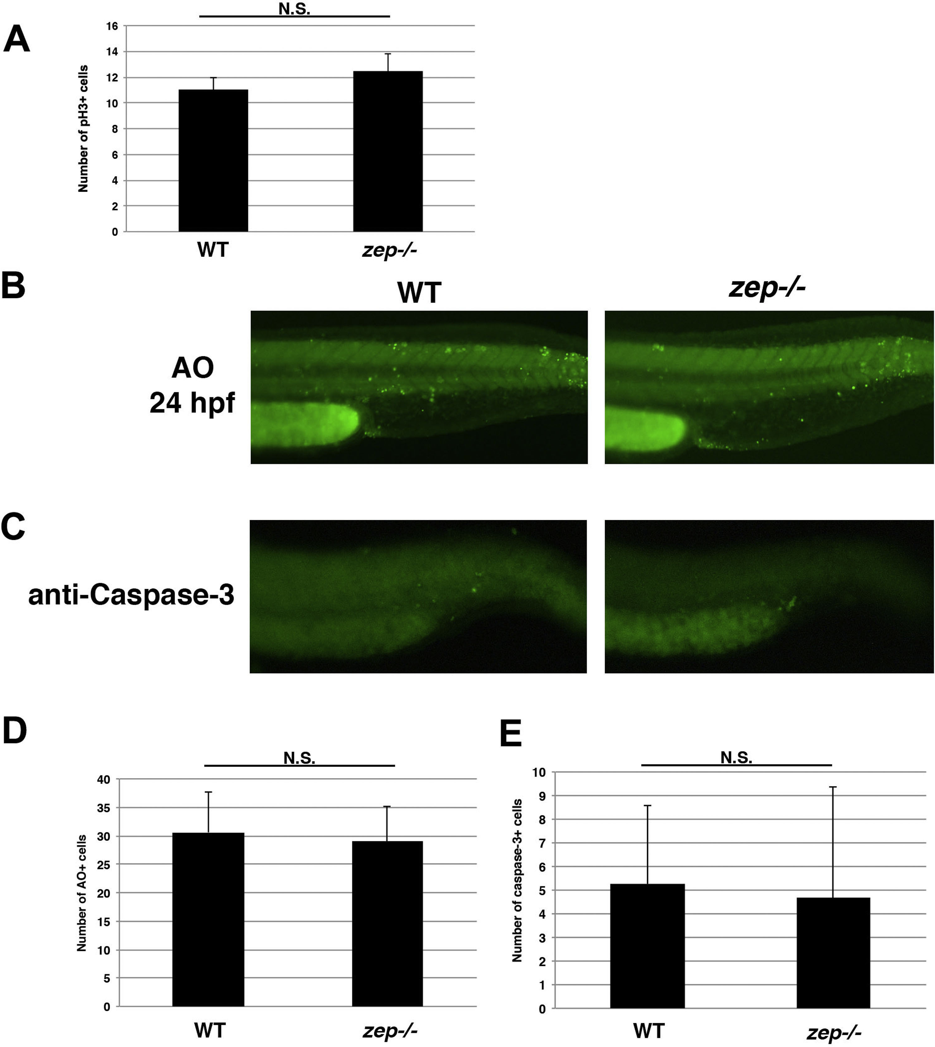
Further assessment of cell proliferation and death inzepmutants. (A) The number of pH3+ cells was quantified in the cervical region of WT and zep embryos at the 24 hpf stage. The quantity of pH3+ cells was not statistically different as assessed by student T-test. (B) Whole mount AO staining and (C) anti-Caspase-3 immunostaining of the tail region in WT and zep embryos revealed similar levels of cell death, which was assessed by student T-tests (D, E) respectively.
Acknowledgments
This image is the copyrighted work of the attributed author or publisher, and
ZFIN has permission only to display this image to its users.
Additional permissions should be obtained from the applicable author or publisher of the image.
Reprinted from Developmental Biology, 428(1), Kroeger, P.T., Drummond, B.E., Miceli, R., McKernan, M., Gerlach, G.F., Marra, A.N., Fox, A., McCampbell, K.K., Leshchiner, I., Rodriguez-Mari, A., BreMiller, R., Thummel, R., Davidson, A.J., Postlethwait, J., Goessling, W., Wingert, R.A., The zebrafish kidney mutant zeppelin reveals that brca2/fancd1 is essential for pronephros development, 148-163, Copyright (2017) with permission from Elsevier. Full text @ Dev. Biol.

