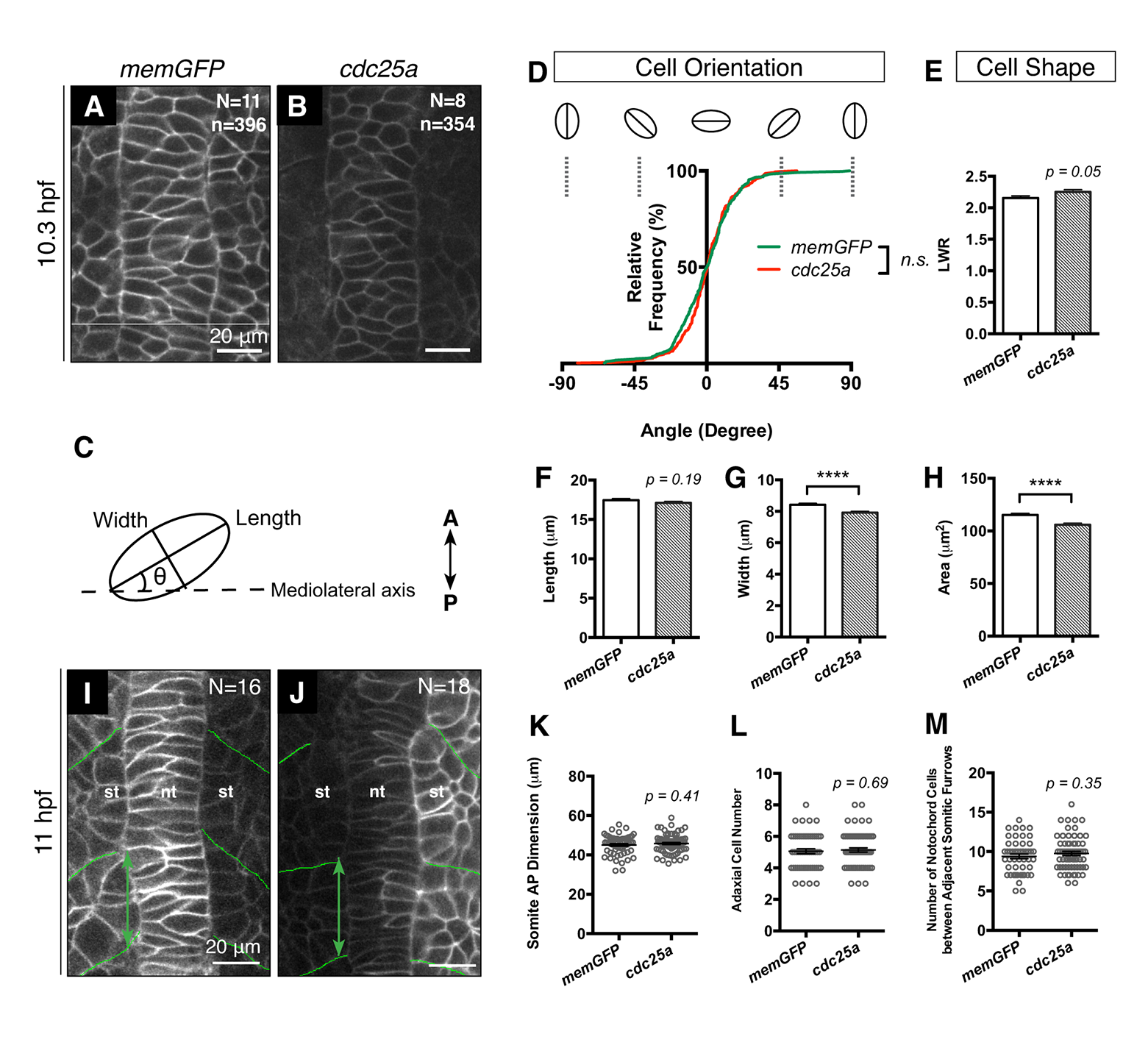
Fig. S9
Effect of cdc25a overexpression in WT on notochord cell size and morphogenesis.
(A and B) Dorsal view of 1-somite stage embryos showing cells labeled with mGFP: control embryos (A) and embryos injected with 25 pg to 50 pg cdc25a mRNA (B) (anterior to the top). (C-H) Analyses of notochord cells’ orientation (D), shape (E), long axis (length, F), short axis (width, G) and size (H) in A and B. (I and J) Confocal image of dorsal mesoderm in 3-somite stage control and cdc25a-overexpressing embryos labeled with mGFP with somite AP dimension illustrated with green arrow and somitic boundaries outlined in green (dorsal view, anterior to the top). (K-M) Quantification of somite AP dimension (K), numbers of adaxial cells (L) and notochord cells (M) in I and J. ****p<0.0001, error bars = SEM.

