Fig. S5
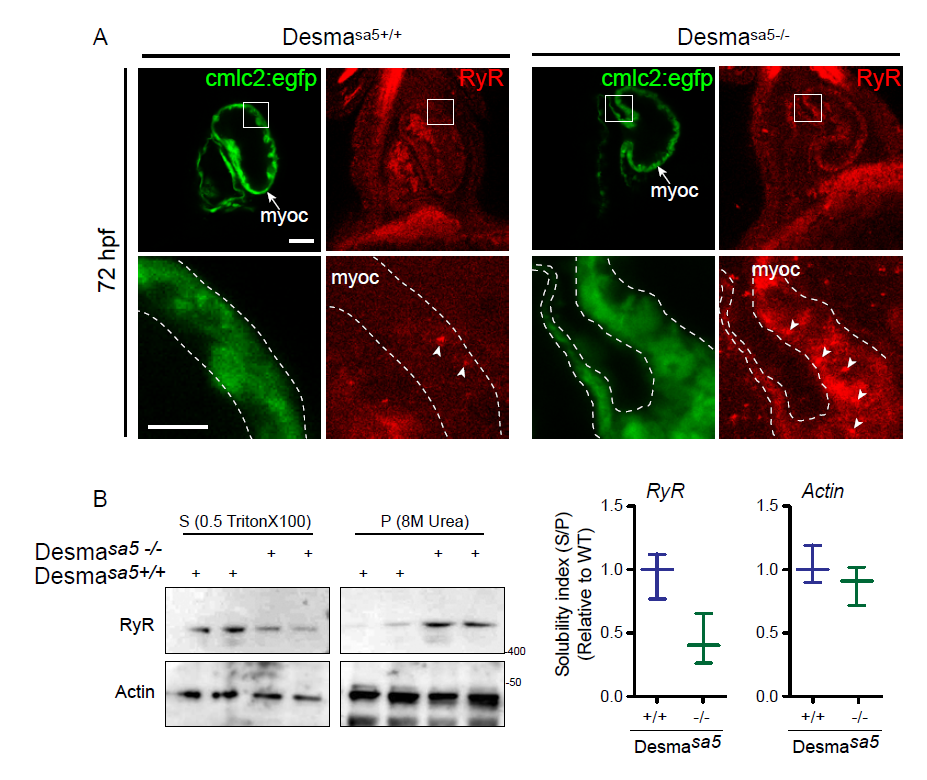
Ryanodine Receptor (RyR) localization is affected in desmasa5+/+ heart (related to Fig.5). (A) RyR localization in desmasa5-/- animals crossed with the myocardium specific Tg(myl7:egfp) (green, myoc, highlighted with the white dotted lines) line compared to controls Tg(myl7:egfp;desmasa5+/+) at 72 hpf. RyR was homogenously distributed (with few small clusters, white arrowheads) in the myocardium in control heart (desmasa5-/-) while it showed important clustering and heterogeneous distribution in the desmasa5+/+ hearts (white arrowheads). Scale bar=30μm, 15μm in the zoom. (B) Confirmation of RyR miss localization in whole fish homogenates by western blot where the major part of RyR protein remains in the X-100 Triton insoluble fraction (Pellet, P) from desmasa5+/+ while RyR is mostly enriched in the X-100 Triton soluble fraction in control samples (desmasa5-/- homogenates). Histograms showing the solubility index (S/P) from 2 independent experiments where actin is the loading control and each homogenate contains a mixture of 80-100 fish. Error bars indicate the standard deviation.

