Image
Figure Caption
Fig. 3
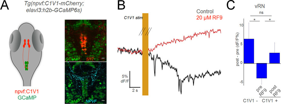
NPVF neurons in the hypothalamus inhibit serotonergic vRN.
(A) Schematic of optogenetic experiments, and overlay of 5HT or NPVF antibody stains on GCaMP+ vRN neurons. (B) Mean response of vRN neurons to optogenetic activation of NPVF neurons, before and after application of gpr147 antagonist RF9. Responses are the mean of all neurons in one example larva (mean ± standard error of the mean (SEM)). (C) Grouped data from C1V1− (5 larvae), and C1V1+ (5 larvae) before and after RF9 application (mean ± SEM). Comparisons are 2-tailed t-tests. *p < 0.05; ns: not significant (p > 0.05).
Acknowledgments
This image is the copyrighted work of the attributed author or publisher, and
ZFIN has permission only to display this image to its users.
Additional permissions should be obtained from the applicable author or publisher of the image.
Full text @ Sci. Rep.

